. 2019 Jan 25;294(4):1095-1103.
doi: 10.1074/jbc.AC118.004905. Epub 2018 Dec 17.

Luminescence-activated nucleotide cyclase regulates spatial and temporal cAMP synthesis

Affiliations

Luminescence-activated nucleotide cyclase regulates spatial and temporal cAMP synthesis

Nyla Naim et al. J Biol Chem. .

Abstract

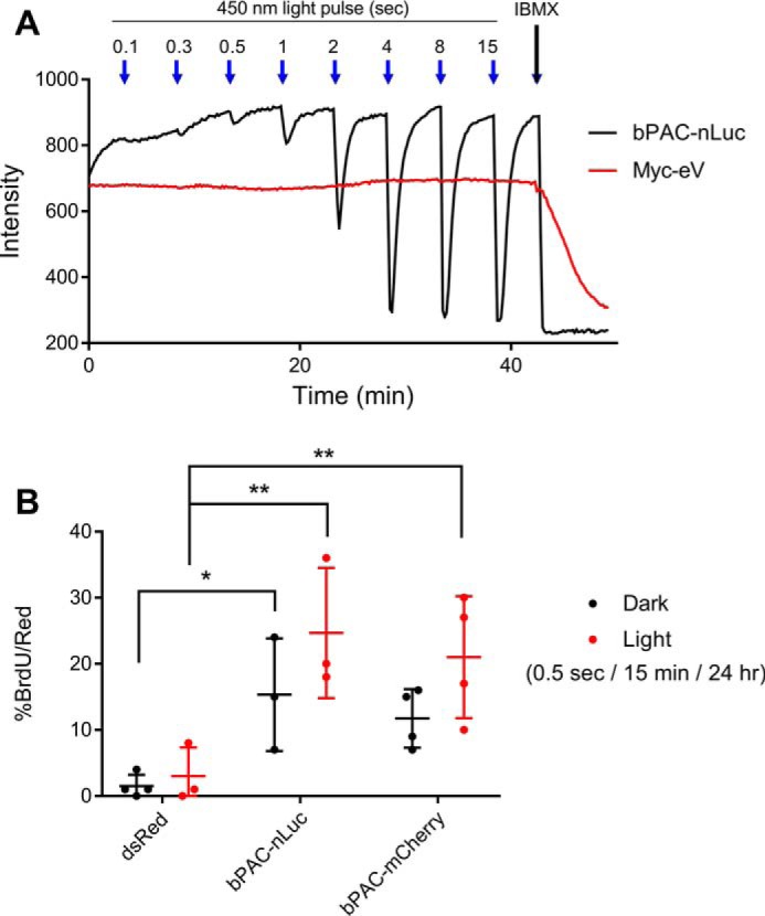
cAMP is a ubiquitous second messenger that regulates cellular proliferation, differentiation, attachment, migration, and several other processes. It has become increasingly evident that tight regulation of cAMP accumulation and localization confers divergent yet specific signaling to downstream pathways. Currently, few tools are available that have sufficient spatial and temporal resolution to study location-biased cAMP signaling. Here, we introduce a new fusion protein consisting of a light-activated adenylyl cyclase (bPAC) and luciferase (nLuc). This construct allows dual activation of cAMP production through temporally precise photostimulation or chronic chemical stimulation that can be fine-tuned to mimic physiological levels and duration of cAMP synthesis to trigger downstream events. By targeting this construct to different compartments, we show that cAMP produced in the cytosol and nucleus stimulates proliferation in thyroid cells. The bPAC-nLuc fusion construct adds a new reagent to the available toolkit to study cAMP-regulated processes in living cells.

Keywords: bPAC; bioluminescence; cell proliferation; compartmentalization; cyclic AMP (cAMP); fluorescence resonance energy transfer (FRET); microdomains; nano-luciferase; optogenetics; second messenger; signaling; thyroid.


Conflict of interest statement

The authors declare that they have no conflicts of interest with the contents of this article

Figures

Figure 1.
Figure 1.
Luminescence-activated cyclase characterization. A, design of dual photo and chemical activator of cAMP production by fusion of bPAC and nLuc. B, transiently transfected HEK293 cells treated with 100 μm h-CTZ luminesce at a maximum of 455 nm (mean ± S.D. of n = 4). C, this effect occurs in a dose-dependent manner (mean ± S.D. of n = 4). D, h-CTZ increases cAMP in HC1 cells expressing bPAC–nLuc in a dose-dependent manner as shown by ELISA analysis (mean ± S.D. of n = 4). E, accumulation of cytosolic cAMP in HEK293 co-expressing H208 and MYR–bPAC–nLuc stimulated with pulses of 4.4 μW/mm2 light (mean ± S.D. of n = 8 cells). F, stimulation with 1:1000 Fz promotes sustained cAMP production well over 30 min (mean ± S.D. of n = 4 cells). RLU, relative luminescence units.
Figure 2.
Figure 2.
Light activation of bPAC–nLuc promotes thyroid cell proliferation. A, real-time cAMP recordings in PCCL3 cells co-expressing red dimerization-dependent cAMP sensor and bPAC–nLuc or myc-empty vector (Myc-eV) control. A decrease in fluorescence represents increased cAMP levels (representative single cell traces shown, 250 μm IBMX shows sensor saturation). B, BrdU incorporation in PCCL3 transiently expressing bPAC–nLuc or bPAC-mCherry upon light exposure (mean ± S.D. of n = 3–4, >500 cells each, two-way analysis of variance with Tukey's multiple comparisons test, *, p < 0.05; **, p < 0.01).
Figure 3.
Figure 3.
Targeted bPAC–nLuc exhibits different levels of activity and effects on thyroid cell proliferation. A, transfected PCCL3 cells stained with anti-Myc (red) and DAPI (blue) expressing bPAC–nLuc constructs throughout the cell (unmodified), NLS, cytosol (NES), and ER. B, PCCL3 co-expressing H208 or NLS-H208 with targeted bPAC–nLuc treated with 1:3000 Fz (mean of >45 cell traces from three independent experiments). The area under the curve from time 0 to 13.5 min normalized between basal and maximum response to 100 μm IBMX (one-way ANOVA with Tukey's multiple comparisons test). C, EdU incorporation in lentiviral-infected stable PCCL3 cell lines expressing targeted bPAC–nLuc constructs treated with 1:1000 Fz-4377. The dotted line shows basal levels of proliferation (mean ± S.D. of n = 5–6, >20,000 cells each, two-way analysis of variance with Dunnett's multiple comparisons test). *, p < 0.05; **, p < 0.01; ****, p < 0.0001.

Similar articles

Cited by

  • CAP1 binds and activates adenylyl cyclase in mammalian cells.
    Zhang X, Pizzoni A, Hong K, Naim N, Qi C, Korkhov V, Altschuler DL. Zhang X, et al. Proc Natl Acad Sci U S A. 2021 Jun 15;118(24):e2024576118. doi: 10.1073/pnas.2024576118. Proc Natl Acad Sci U S A. 2021. PMID: 34099549 Free PMC article.
  • Engineered BRET-Based Biologic Light Sources Enable Spatiotemporal Control over Diverse Optogenetic Systems.
    Parag-Sharma K, O'Banion CP, Henry EC, Musicant AM, Cleveland JL, Lawrence DS, Amelio AL. Parag-Sharma K, et al. ACS Synth Biol. 2020 Jan 17;9(1):1-9. doi: 10.1021/acssynbio.9b00277. Epub 2019 Dec 17. ACS Synth Biol. 2020. PMID: 31834783 Free PMC article.
  • Whole-Cell cAMP and PKA Activity are Epiphenomena, Nanodomain Signaling Matters.
    Bers DM, Xiang YK, Zaccolo M. Bers DM, et al. Physiology (Bethesda). 2019 Jul 1;34(4):240-249. doi: 10.1152/physiol.00002.2019. Physiology (Bethesda). 2019. PMID: 31165682 Free PMC article. Review.
  • Neurophotonic tools for microscopic measurements and manipulation: status report.
    Abdelfattah AS, Ahuja S, Akkin T, Allu SR, Brake J, Boas DA, Buckley EM, Campbell RE, Chen AI, Cheng X, Čižmár T, Costantini I, De Vittorio M, Devor A, Doran PR, El Khatib M, Emiliani V, Fomin-Thunemann N, Fainman Y, Fernandez-Alfonso T, Ferri CGL, Gilad A, Han X, Harris A, Hillman EMC, Hochgeschwender U, Holt MG, Ji N, Kılıç K, Lake EMR, Li L, Li T, Mächler P, Miller EW, Mesquita RC, Nadella KMNS, Nägerl UV, Nasu Y, Nimmerjahn A, Ondráčková P, Pavone FS, Perez Campos C, Peterka DS, Pisano F, Pisanello F, Puppo F, Sabatini BL, Sadegh S, Sakadzic S, Shoham S, Shroff SN, Silver RA, Sims RR, Smith SL, Srinivasan VJ, Thunemann M, Tian L, Tian L, Troxler T, Valera A, Vaziri A, Vinogradov SA, Vitale F, Wang LV, Uhlířová H, Xu C, Yang C, Yang MH, Yellen G, Yizhar O, Zhao Y. Abdelfattah AS, et al. Neurophotonics. 2022 Jan;9(Suppl 1):013001. doi: 10.1117/1.NPh.9.S1.013001. Epub 2022 Apr 27. Neurophotonics. 2022. PMID: 35493335 Free PMC article.
  • Dual Activation of cAMP Production Through Photostimulation or Chemical Stimulation.
    Naim N, Reece JM, Zhang X, Altschuler DL. Naim N, et al. Methods Mol Biol. 2020;2173:201-216. doi: 10.1007/978-1-0716-0755-8_14. Methods Mol Biol. 2020. PMID: 32651920 Free PMC article.

References

    1. Sutherland E. W., and Rall T. W. (1958) Fractionation and characterization of a cyclic adenine ribonucleotide formed by tissue particles. J. Biol. Chem. 232, 1077–1091 - PubMed
    1. Fajardo A. M., Piazza G. A., and Tinsley H. N. (2014) The role of cyclic nucleotide signaling pathways in cancer: targets for prevention and treatment. Cancers 6, 436–458 10.3390/cancers6010436 - DOI - PMC - PubMed
    1. Perera R. K., and Nikolaev V. O. (2013) Compartmentation of cAMP signalling in cardiomyocytes in health and disease. Acta Physiol. 207, 650–662 10.1111/apha.12077 - DOI - PubMed
    1. Berdeaux R., and Stewart R. (2012) cAMP signaling in skeletal muscle adaptation: hypertrophy, metabolism, and regeneration. Am. J. Physiol. Endocrinol. Metab. 303, E1–E17 10.1152/ajpendo.00555.2011 - DOI - PMC - PubMed
    1. Miller R. A., Chu Q., Xie J., Foretz M., Viollet B., and Birnbaum M. J. (2013) Biguanides suppress hepatic glucagon signalling by decreasing production of cyclic AMP. Nature 494, 256–260 10.1038/nature11808 - DOI - PMC - PubMed

Publication types