Review
. 2018 Dec 3:9:2901.
doi: 10.3389/fmicb.2018.02901. eCollection 2018.

Host Variability in NTM Disease: Implications for Research Needs

Affiliations
Review

Host Variability in NTM Disease: Implications for Research Needs

Colin Swenson et al. Front Microbiol. .

Abstract

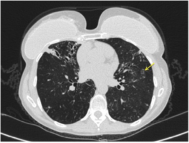
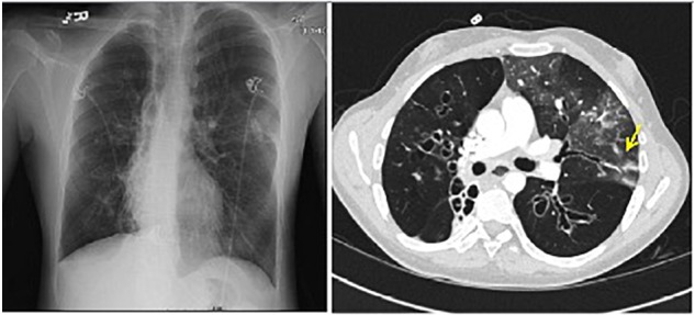
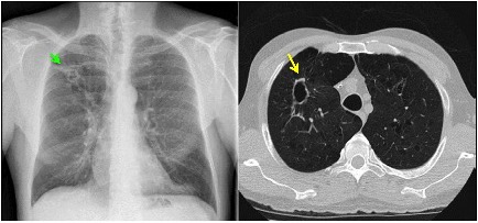
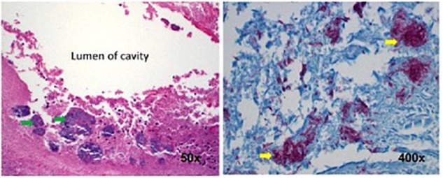
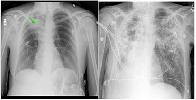
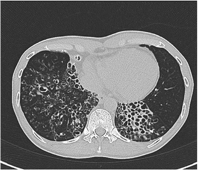
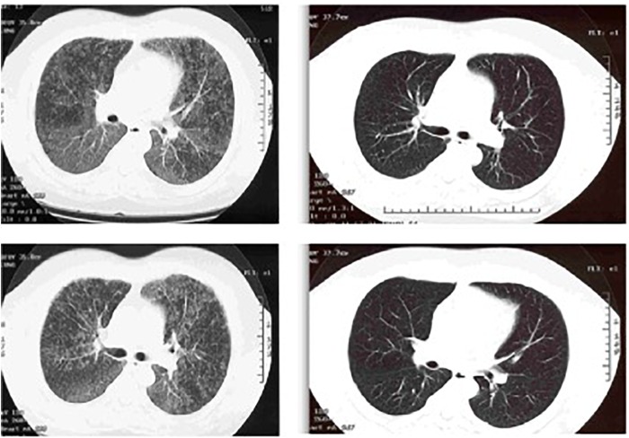
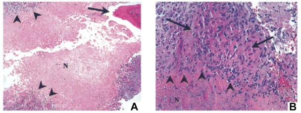
Non-tuberculous mycobacteria (NTM) are ubiquitous environmental organisms that may cause opportunistic infections in susceptible hosts. Lung infections in immunocompetent persons with structural lung disease are most common, while disseminated disease occurs primarily in immunocompromised individuals. Human disease caused by certain species, such as Mycobacterium avium complex, Mycobacterium abscessus, and Mycobacterium kansasii, is increasing in incidence and varies by geographic distribution. The spectrum of NTM disease varies widely in presentation and clinical outcome, but certain patterns can be organized into clinical phenotypes. Treatment options are limited, lengthy, and often toxic. The purpose of this case-based review is to provide non-clinician scientists with a better understanding of human NTM disease with an aim to stimulate more research and development.

Keywords: COPD; bronchiectasis; cystic fibrosis; disseminated; non-tuberculous mycobacteria; osteomyelitis; pulmonary.


Figures

FIGURE 1
FIGURE 1
Axial CT chest image demonstrating bronchiectasis and mucoid impaction in the right middle lobe. The tree-in-bud nodularity in the left lung (yellow arrow).
FIGURE 2
FIGURE 2
Left: chest radiograph demonstrating cystic bronchiectasis. Right: CT image of chest showing a left upper lobe infiltrate on a background of cystic bronchiectasis (yellow arrow).
FIGURE 3
FIGURE 3
Left: chest radiograph demonstrating a right upper lobe cavitary lesion on a background of emphysema (green arrow). Right: axial CT image demonstrating the same cavitary lesion in the right lung (yellow arrow). Reprinted with permission of the American Thoracic Society. Copyright© 2018 American Thoracic Society. Fennelly et al. (2016) is an official journal of the American Thoracic Society.
FIGURE 4
FIGURE 4
Histopathology of the surgical specimen (using hematoxylin and eosin stains) demonstrating aggregates of bacteria (left, green arrows) along the inner wall of the lung cavity, with many AFB seen at higher power using Ziehl Nissen stain (right, yellow arrows). Reprinted with permission of the American Thoracic Society. Copyright© 2018 American Thoracic Society. Fennelly et al. (2016) is an official journal of the American Thoracic Society.
FIGURE 5
FIGURE 5
Left: chest radiograph showing right upper lobe consolidation with central cavitary changes (green arrow) in a woman with COPD and pulmonary NTM disease. Right: chest radiograph of same subject demonstrating marked progression of pulmonary NTM disease, including likely spillage of the previous right upper lobe cavitary lesion.
FIGURE 6
FIGURE 6
CT chest image showing cystic bronchiectasis in the right and left lower lobes.
FIGURE 7
FIGURE 7
Left: CT chest showing diffuse ground glass centrilobular nodules and mosaic attenuation. Right: post-treatment images demonstrating resolution of the prior findings.
FIGURE 8
FIGURE 8
Bone biopsy specimen (T9 vertebra) of patient 1 showing necrotizing granulomas. (A) Area of necrotic bone is shown (arrow; hematoxylin and eosin stain; original magnification, ×100). Surrounding the area of caseous necrosis (N) is a rim of granulomatous tissue reaction (arrowheads). (B) At higher magnification (hematoxylin and eosin stain; magnification, ×400), the granulomas are principally composed of macrophages (area between the arrows) and lymphocytes (arrowheads). Acid-fast and fungal stains showed negative results, but culture was positive for Mycobacterium abscessus. Reprinted with permission of Oxford University Press; Chan et al. (2001).

References

    1. Adjemian J., Olivier K. N., Prevots D. R. (2014a). Nontuberculous mycobacteria among patients with cystic fibrosis in the United States: screening practices and environmental risk. Am. J. Respir. Crit. Care Med. 190 581–586. 10.1164/rccm.201405-0884OC - DOI - PMC - PubMed
    1. Adjemian J., Olivier K. N., Prevots D. R. (2018). Epidemiology of pulmonary Nontuberculous Mycobacterial sputum positivity in patients with cystic fibrosis in the United States, 2010-2014. Ann. Am. Thorac. Soc. 15 817–826. 10.1513/AnnalsATS.201709-727OC - DOI - PMC - PubMed
    1. Adjemian J., Prevots D. R., Gallagher J., Heap K., Gupta R., Griffith D. (2014b). Lack of adherence to evidence-based treatment guidelines for nontuberculous mycobacterial lung disease. Ann. Am. Thorac. Soc. 11 9–16. 10.1513/AnnalsATS.201304-085OC - DOI - PMC - PubMed
    1. Adjemian J., Seitz A. E., Holland S. M., Prevots D. R. (2012). Prevalence of nontuberculous mycobacterial lung disease in U.S. Medicare beneficiaries. Am. J. Respir. Crit. Care Med. 185 881–886. 10.1164/rccm.201111-2016OC - DOI - PMC - PubMed
    1. Boyle D. P., Zembower T. R., Qi C. (2016). Relapse versus Reinfection of Mycobacterium avium complex Pulmonary disease. Patient characteristics and macrolide susceptibility. Ann. Am. Thorac. Soc. 13 1956–1961. 10.1513/AnnalsATS.201605-344BC - DOI - PubMed

LinkOut - more resources