. 2018 Dec 3:9:2918.
doi: 10.3389/fmicb.2018.02918. eCollection 2018.

Novel Widespread Marine Oomycetes Parasitising Diatoms, Including the Toxic Genus Pseudo-nitzschia: Genetic, Morphological, and Ecological Characterisation

Affiliations

Novel Widespread Marine Oomycetes Parasitising Diatoms, Including the Toxic Genus Pseudo-nitzschia: Genetic, Morphological, and Ecological Characterisation

Andrea Garvetto et al. Front Microbiol. .

Abstract

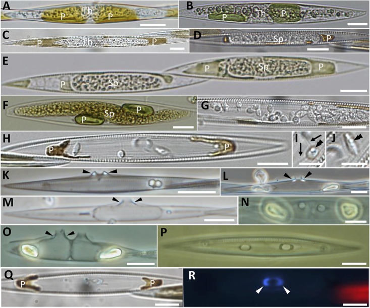
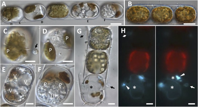
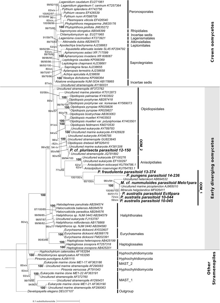
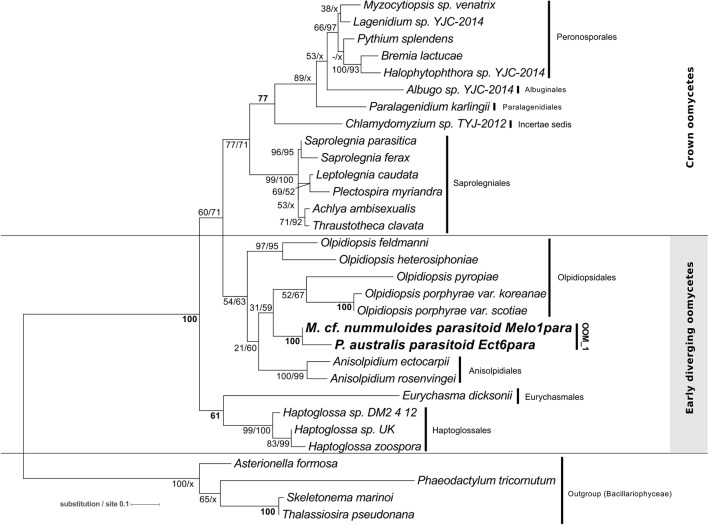
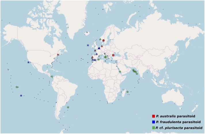
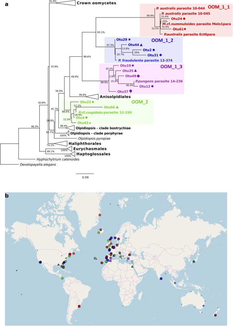
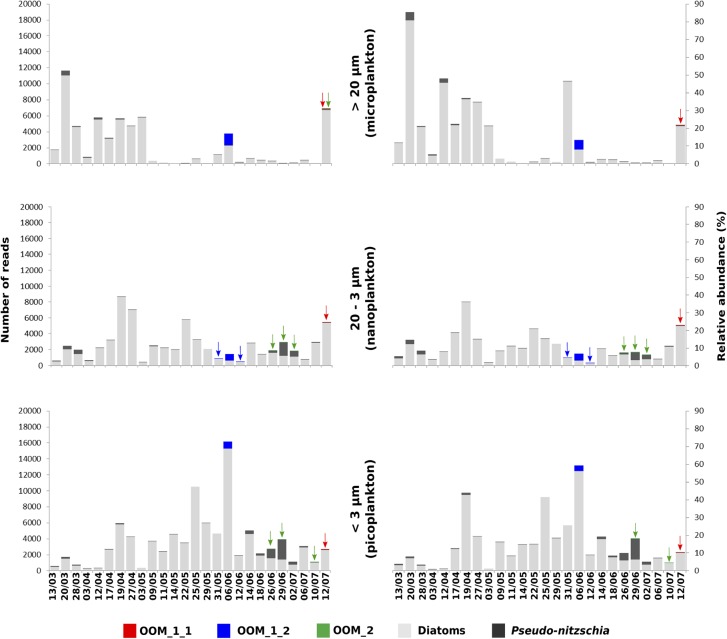
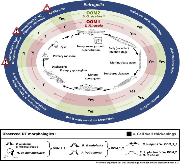
Parasites are key drivers of phytoplankton bloom dynamics and related aquatic ecosystem processes. Yet, the dearth of morphological and molecular information hinders the assessment of their diversity and ecological role. Using single-cell techniques, we characterise morphologically and molecularly, intracellular parasitoids infecting four potentially toxin-producing Pseudo-nitzschia and one Melosira species on the North Atlantic coast. These sequences define two, morphologically indistinguishable clades within the phylum Oomycota, related to the genera of algal parasites Anisolpidium and Olpidiopsis and the diatom parasitoid species Miracula helgolandica. Our morphological data are insufficient to attribute either clade to the still unsequenced genus Ectrogella; hence it is proposed to name the clades OOM_1 and OOM_2. A screening of global databases of the barcode regions V4 and V9 of the 18S rDNA demonstrate the presence of these parasitoids beyond the North Atlantic coastal region. During a biweekly metabarcoding survey (Concarneau Bay, France), reads associated with one sequenced parasitoid coincided with the decline of Cerataulina pelagica bloom, whilst the other parasitoids co-occurred at low abundance with Pseudo-nitzschia. Our data highlight a complex and unexplored diversity of the oomycete parasitoids of diatoms and calls for the investigation of their phenology, evolution, and potential contribution in controlling their host spatial-temporal dynamics.

Keywords: Pseudo-nitzschia; diatoms; marine oomycetes; metabarcoding; plankton parasites; single-cell analysis.


Figures

FIGURE 1
FIGURE 1
Morphological characterisation of the novel oomycete parasitoids of Pseudo-nitzschia described in this study. (A–C) Unwalled, increasingly granular thalli developing in Pseudo-nitzschia australis (A,B) and Pseudo-nitzschia cf. pungens/multiseries cells (C); thalli are progressively pushing apart the degraded, yet still pigmented plastids. (D–F) Holocarpic syncytia maturing into sporangia, in Pseudo-nitzschia cf. pungens/multiseries (D,E) and Pseudo-nitzschia australis (F). (G–J) Zoospore release in Pseudo-nitzschia cf. pungens/multiseries. Zoospores bear two flagella (arrows in I), a single refractive globule (double arrowheads in I and J) and their shape varies from almost spherical (I) to oblong (J). (K–R) Empty sporangia. Arrowheads point to thickenings in the discharge tube. (K) Pseudo-nitzschia pungens/multiseries, valve view. (L) Pseudo-nitzschia australis, valve view. (M) Empty sporangium within Pseudo-nitzschia fraudulenta, valve view. (N–P) Two empty sporangia within a Pseudo-nitzschia fraudulenta frustule, girdle view (N) and valve view (O), and Pseudo-nitzschia pungens/multiseries, girdle view (P). (Q,R) Pseudo-nitzschia cf. pungens/multiseries, girdle view, under DIC (Q) and epifluorescence microscopy (R, Calcofluor-White staining), to highlight the thickening of the discharge tube. Scale bars = 10 μm except in I,J (5 μm). Samples in (B,E,F) were Lugol’s iodine fixed. (B) Refers to the sequenced cell P. australis parasitoid 10-044. Th, thallus; Sp, sporangium; P, plastid.
FIGURE 2
FIGURE 2
Morphological characterisation of a novel oomycete parasitoid of Melosira cf. nummuloides. (A,B). Highly infected diatom chain (A), compared to a healthy one (B). (C) Early stage of infection, with a spore (arrow) bearing refractive globules, encysted on a diatom frustule. (D) Unwalled thallus developing within the host cell, the once numerous small plate-like plastids are collapsed in two olive-green masses. (E) Highly vacuolised parasitic thallus filling the host cell almost entirely. (F) Mature spherical multinucleate thallus filling most of the host cell, the disrupted plastids are represented by two brown masses. (G,H) Bright field (G) and epifluorescence (H, Calcofluor-White staining) microscopy of an uninfected cell (top) and a dead cell bearing one empty sporangium (bottom, asterisk) and an encysted spore (arrow). At a different focal plan (H, right panel), a second smaller parasitic sporangium was discernible (top asterisk). Sporangial wall thickenings in correspondence of the discharge tube were also visible (arrowheads). (A,C–F) Refers to the single infected colony sequenced to obtain M. cf. nummuloides parasitoid Melo1para sequence. P, plastids; V, vacuole; Th, thallus. Scale bars: 10 μm in (A,B); 5 μm from (C–H).
FIGURE 3
FIGURE 3
Maximum likelihood phylogenetic tree reconstruction based on partial 18S rDNA genes of novel diatom parasitoids within the Oomycota (Stramenopila), inferred from 81 sequences on 1,206 positions. New sequences are in bold and they are named according to the host species and the sample ID within the new clades OOM_1 and OOM_2 (details in Table 1). Bootstrap values (1,000 replicates for ML and NJ, 100 for MP) are shown at each node as a percentage for the three computational methods tested, with the following order: maximum likelihood, neighbour joining and maximum parsimony. X, node not supported; –, bootstrap support <50%. Bold numbers indicate agreement on the same bootstrap value for the three methods. Refer to the alignment in Supplementary Data Sheet S2.
FIGURE 4
FIGURE 4
Maximum-likelihood phylogenetic tree reconstruction using 31 concatenated amino acid sequences for the mitochondrial genetic markers cox1 (194 positions) and cox2 (185 positions) of the parasitoid of Melosira cf. nummuloides (in bold) within the oomycetes (Stramenopila). The cox2 amino acid sequence of the parasitoid of P. australis Ect6para (in bold) was also included. Four diatoms (Stramenopila, Bacillariophyceae) were defined as outgroup. Bootstrap values (1,000 replicates) are shown at each node for ML and NJ methods, respectively. Bold numbers indicate agreement on the same bootstrap value for both methods. Refer to the alignment in Supplementary Data Sheet S3.
FIGURE 5
FIGURE 5
Worldwide screening for the presence of Pseudo-nitzschia oomycete parasitoid barcodes. The metabarcode databases Ocean Sampling Day (triangle), BioMarKs (circle), and TARA (star) were searched for the presence of >10 paired reads over 99% identical to the V4 or V9 region of the 18S rDNA of the parasitoids P. cf. plurisecta 12-150 (green) within OOM_2; P. fraudulenta 13-374 (blue), P. australis 10-044/10-045 (red) within OOM_1. Small black symbols show the absence of reads matching the searched organisms in the relative database.
FIGURE 6
FIGURE 6
Diversity and distribution of OOM_1 and OOM_2 related OTUs based on public SRA marine barcoding datasets. The colour code identifies each one of the four new clades of diatom parasitoids OOM_1_1 (red), OOM_1_2 (blue), OOM_1_3 (purple) and OOM_2 (green). (a) Maximum Likelihood tree of 15 18S rDNA V4 OTUs retrieved by the MOULINETTE Pipeline. Refer to the alignment in Supplementary Data Sheet S4. (b) Global distribution of each detected OTU based on GPS coordinates of matching SRA runs. Within-clade symbols are attributed to different OTUs.
FIGURE 7
FIGURE 7
Phenology of the parasitoid of P. cf. plurisecta 12-150 (OOM_2, green) and of putative parasitoids within the sub-clades OOM_1_1 (red) and OOM_1_2 (blue), in relation to the diatom community during the spring/summer 2012 in the Bay of Concarneau (France), described by metabarcoding survey. The three left hand-side histograms show the total abundance of reads on the y-axis, whilst the ones on the right hand side show the relative abundances as a percentage of the total eukaryotic community. The size fractions for microplankton (top, >20 μm), nanoplankton (middle, 20–3 μm) and picoplankton (bottom, 3–0.2 μm) are indicated in the middle. Pseudo-nitzschia OTUs are in dark grey, other diatom OTUs in light grey. Sampling dates are shown on the bottom x-axis.
FIGURE 8
FIGURE 8
Mapping observed OOM_1 and Miracula helgolandica (Buaya et al., 2017) and OOM_2 and Olpidiopsis drebesii (Buaya et al., 2017) life stages against Pseudo-nitzschia pungens oomycete parasitoid life cycle (Hanic et al., 2009) and the defining morphological criteria for the Ectrogella genus (Zopf, 1884; Scherffel, 1925). The development cycle of the oomycete parasitoid of P. pungens described by Hanic et al. (2009) is reproduced in the inner ellipse. The key morphological criteria defining the genus Ectrogella Zopf emend. Scherffel are highlighted in the external dark blue ellipse. The warning signs highlight contradictions between the original descriptions of the type species given by Zopf and Scherffel. The intercalary red and green ellipses highlight the congruence of each criterion (Yes) or lack thereof (?!, when morphology unknown or conflicting) between these original Ectrogella descriptions and the morphology of the OOM_1 and OOM_2 clades, respectively. The box at the bottom summarises the different DT morphologies as observed in different diatom hosts. Black colour highlights the cell wall thickenings (“forcing apparatus” sensu Sparrow, 1960).

References

    1. Alster A., Zohary T. (2007). Interactions between the bloom-forming dinoflagellate Peridinium gatunense and the chytrid fungus Phlyctochytrium sp. Hydrobiologia 578 131–139. 10.1007/s10750-006-0439-y - DOI
    1. Altschul S. F., Gish W., Miller W., Myers E. W., Lipman D. J. (1990). Basic local alignment search tool. J. Mol. Biol. 215 403–410. 10.1016/S0022-2836(05)80360-2 - DOI - PubMed
    1. Beakes G. W., Glockling S. L., Sekimoto S. (2012). The evolutionary phylogeny of the oomycete “fungi.”. Protoplasma 249 3–19. 10.1016/j.ympev.2015.03.003 - DOI - PubMed
    1. Beakes G. W., Honda D., Thines M. (2014). “Systematics of the straminipila: labyrinthulomycota, hyphochytriomycota, and oomycota,” in Systematics and Evolution, 2nd Edn, eds McLaughlin D. J., Spatafora J. W. (Berlin: Springer-Verlag; ), 39–97.
    1. Beakes G. W., Thines M. (2016). “Hyphochytriomycota and oomycota,” in Handbook of the Protists, eds Archibald J. M., Simpson A. G. B., Slamovits C. (Cham: Springer International Publishing; ), 3–71.

LinkOut - more resources