. 2018 Dec 3:9:2830.
doi: 10.3389/fimmu.2018.02830. eCollection 2018.

The Adhesion G Protein-Coupled Receptor GPR97/ ADGRG3 Is Expressed in Human Granulocytes and Triggers Antimicrobial Effector Functions

Affiliations

The Adhesion G Protein-Coupled Receptor GPR97/ ADGRG3 Is Expressed in Human Granulocytes and Triggers Antimicrobial Effector Functions

Cheng-Chih Hsiao et al. Front Immunol. .

Abstract

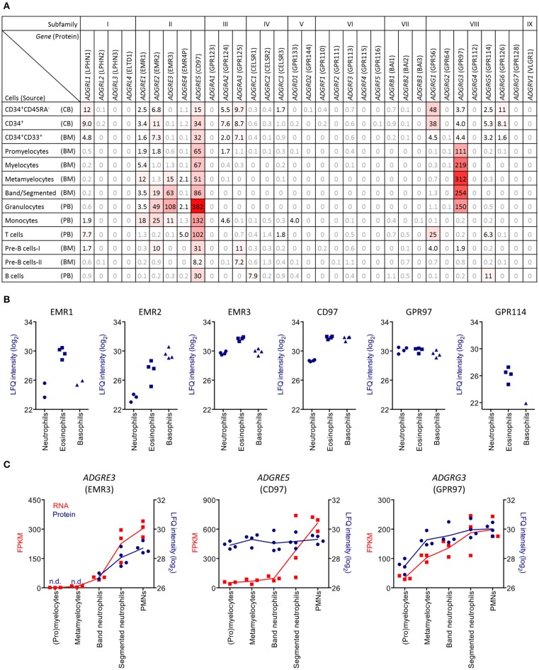
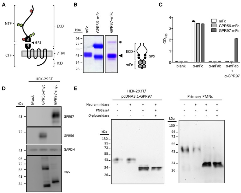
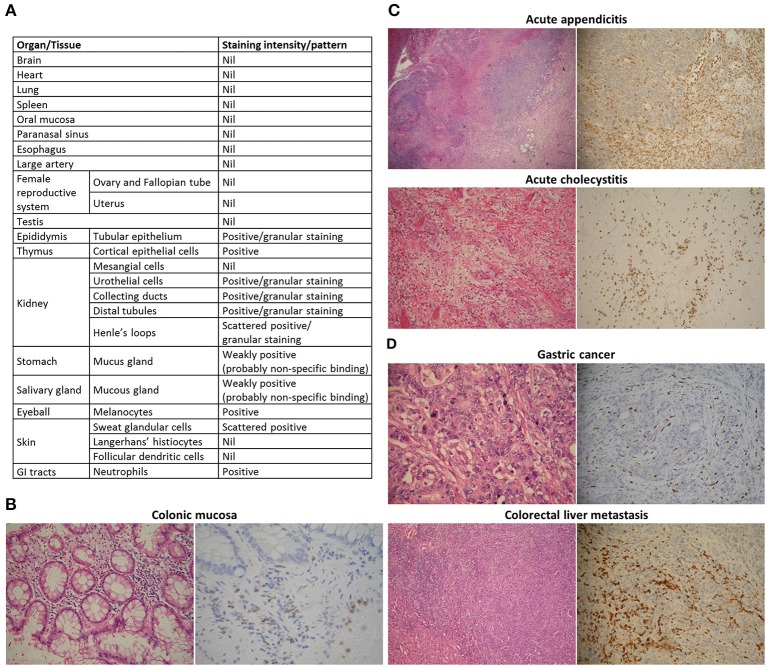
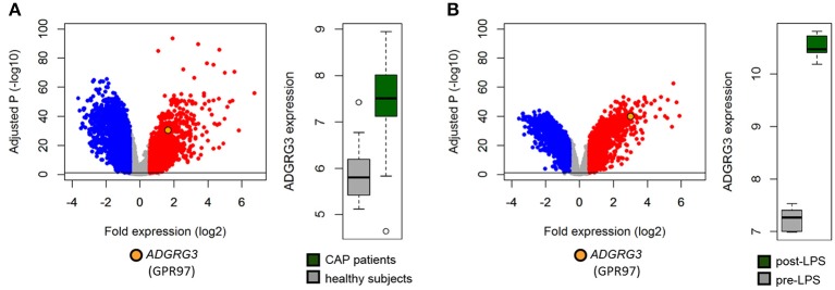
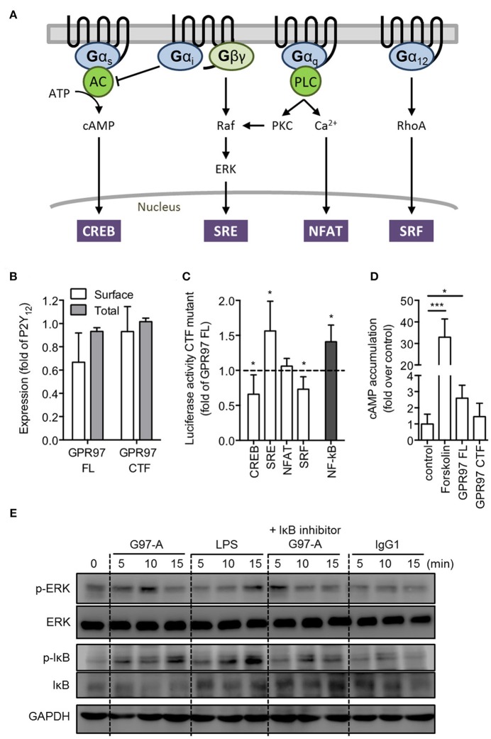
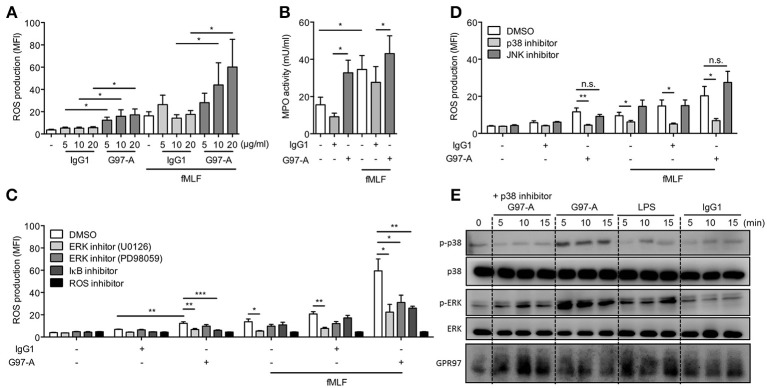
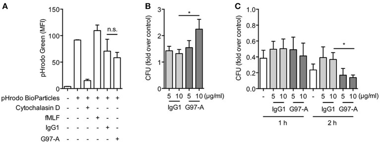
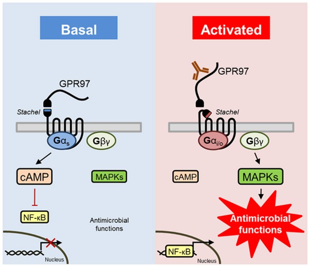
The adhesion family of G protein-coupled receptors (aGPCRs) comprises 33 members in human, several of which are distinctly expressed and functionally involved in polymorphonuclear cells (PMNs). As former work indicated the possible presence of the aGPCR GPR97 in granulocytes, we studied its cellular distribution, molecular structure, signal transduction, and biological function in PMNs. RNA sequencing and mass-spectrometry revealed abundant RNA and protein expression of ADGRG3/GPR97 in granulocyte precursors and terminally differentiated neutrophilic, eosinophilic, and basophilic granulocytes. Using a newly generated GPR97-specific monoclonal antibody, we confirmed that endogenous GPR97 is a proteolytically processed, dichotomous, N-glycosylated receptor. GPR97 was detected in tissue-infiltrating PMNs and upregulated during systemic inflammation. Antibody ligation of GPR97 increased neutrophil reactive oxygen species production and proteolytic enzyme activity, which is accompanied by an increase in mitogen-activated protein kinases and IκBα phosphorylation. In-depth analysis of the GPR97 signaling cascade revealed a possible switch from basal Gαs/cAMP-mediated signal transduction to a Gαi-induced reduction in cAMP levels upon mutation-induced activation of the receptor, in combination with an increase in downstream effectors of Gβγ, such as SRE and NF-κB. Finally, ligation of GPR97 increased the bacteria uptake and killing activity of neutrophils. We conclude that the specific presence of GPR97 regulates antimicrobial activity in human granulocytes.

Keywords: G-protein signaling; adhesion GPCR; antimicrobial activity; granulocytes; inflammation; monoclonal antibody.


Figures

Figure 1
Figure 1
PMNs specifically express ADGRG3 (GPR97). (A) RNA sequencing data showing aGPCR expression in sorted cord blood (CB), bone marrow (BM), and peripheral blood (PB) cell populations, derived from Maiga et al. (25). Shown are aGPCR with reads per kilobase per million mapped reads (RPKM) >1.5. (B) Mass spectrometry data showing protein expression of the aGPCRs EMR1, EMR2, EMR3, CD97, GPR97, and GPR114 in granulocyte populations, including neutrophils, eosinophils, and basophils, derived from Rieckmann et al. (26). (C) Linked RNA sequencing and mass spectrometry data showing expression of the aGPCRs ADGRE3 (EMR3), ADGRE5 (CD97), and ADGRG3 (GPR97) in precursor and mature granulocytes. FPKM, fragments per kilobase per million mapped reads; LFQ, label-free quantification.
Figure 2
Figure 2
GPR97 is a dichotomous receptor. (A) Schematic presentation of GPR97 architecture with the N-terminal and C-terminal fragment (NTF and CTF), linked by the (GPCR autoproteolysis-inducing) GAIN domain with a GPCR proteolysis site (GPS), predicted to facilitate autoproteolysis of the receptor. Alternatively, the protein layout of GPR97 is marked by a three-partite structure consisting of an extracellular domain (ECD), a seven transmembrane (7TM) domain, and an intracellular domain (ICD). N- and O-glycosylation sites were predicted by NetNGlyc and NetOGlyc (http://www.cbs.dtu.dk/services/) and are indicated by green pentagons and red circles, respectively. (B) SDS-PAGE analysis of the GPR97-ECD–mFc protein (schematically provided to the right) used for mAb production. The molecular weight of GPR97-NTF is ~43 kDa (indicated by arrowhead), similar to the size of mFc. A minor fraction of uncleaved GPR97-ECD–mFc was expressed as a ~80-kDa band (indicated by asterisk). mFc and GPR56-ECD–mFc protein were included as controls. (C) ELISA demonstrating the specificity of the anti-GPR97 mAb G97-A. ELISA plates coated with mFc, GPR56-ECD–mFc, or GPR97-ECD–mFc protein were probed with Abs directed against mouse IgG Fc, mouse IgG Fab, or GPR97 (G97-A), followed by color development of tetramethylbenzidine substrate. (D) Western blot analysis of GPR97 receptor expressed in HEK-293T cells transiently expressing GPR97–myc. Data show that GPR97 is proteolytically cleaved into a ~43-kDa NTF and a ~38-kDa CTF, detected by anti-GPR97 (G97-A) and anti-myc mAbs, respectively. HEK-293T cells expressing GPR56–myc protein were included as controls. (E) Total cell lysates of HEK-293T cells transiently expressing GPR97 and primary PMNs were treated with or without neuraminidase, PNGaseF, and O-glycosidase and probed with G97-A mAb. As indicated, GPR97 protein was heavily decorated by N-linked glycosylation.
Figure 3
Figure 3
Tissue-infiltrating PMNs express GPR97. Tissue reactivity of the anti-GPR97 mAb G97-A on formalin-fixed and paraffin-embedded human tissues. (A) Summary of GPR97 expression in human organs. (BD) Left: hematoxylin and eosin staining. Right: immunohistochemical staining. G97-A-stained neutrophils in (B) colonic mucosa (400 × ), (C) acute appendicitis and acute cholecystitis (both 200 × ), and (D) gastric adenocarcinoma and colorectal liver metastasis (both 200 × ).
Figure 4
Figure 4
Inflammation enhances ADGRG3 expression. Elevated whole blood expression of GPR97 was detected in (A) critically ill community-acquired pneumonia patients (n = 101), relative to healthy subjects (n = 42), and (B) healthy volunteers at 4 h of endotoxemia (n = 7) initiated by administration 4 ng/kg E. coli-derived LPS, intravenously, relative to pre-LPS administration (n = 7). Red dots denote significant genes with fold expression >1.5. Blue dots denote significant genes with fold expression < -1.5. –log10 (BH) p, negative log-transformed Benjamini-Hochberg adjusted probabilities. Horizontal lines denote multiple comparison-adjusted probability of 0.05.
Figure 5
Figure 5
GPR97 mutation suppresses cAMP production and increases NF-κB activity enhances. (A) Schematic presentation of GPCR signaling pathways [adapted from Cheng et al. (41)]. Gαs-coupled receptors activate adenylate cyclase (AC), leading to cAMP accumulation. Gαi-coupled receptors inhibit AC, and their Gβγ subunits activate MAPK, such as ERK. Gαq-coupled receptors activate phospholipase C (PLC) to increase intracellular calcium concentration as well as activate protein kinase C (PKC), which results in Raf kinase activation of the MAPK pathway. Gα12-coupled receptors activate the small GTPase RhoA. Downstream reporters are cAMP response element-binding protein (CREB), serum response element (SRE), nuclear factor of activated T cells (NFAT), and serum response factor (SRF). (B) Surface and total cell expression of full-length (FL) and CTF-only mutant GPR97 in COS-7 cells. OD values are given as the percentage of human P2Y12, which served as a positive control (basal expression: empty vector: 0.01 ± 0.02 OD492nm; P2Y12: 1.304 ± 0.236 OD492nm). (C) Activity levels of GPR97–CTF in luciferase reporter gene assays on HEK-293T transfected cells. Provided is the percentage of the signal in relation to full-length GPR97 and after normalization to control in the given luciferase activity (full-length activity – fold over control: CREB: 1.31 ± 0.55; SRE: 1.12 ± 0.15; NFAT: 0.88 ± 0.22; SRF: 1.34 ± 0.57; NF-κB: 1.15 ± 0.17). (D) cAMP accumulation in COS-7 cells treated with forskolin or transfected with full-length and mutant GPR97. Data are shown as fold over control. (E) Isolated peripheral blood PMNs were incubated with control IgG1 or G97-A mAb (10 μg/ml) at 4°C, followed by a treatment with goat-anti mouse F(ab')2 at 4°C, and then shifted cells to 37°C for different time points. LPS treatment (100 ng/ml) served as positive control, and NF-κB signaling was inhibited with IκBα inhibitor (Bay 11–7082, 5 μM). Phospho- and total protein were detected by Western blot analysis. All data are means ± SEM of 4 independent experiments performed in triplicate. *p < 0.05; ***p < 0.001.
Figure 6
Figure 6
Antibody ligation of GPR97 enhances antimicrobial mediator production of PMNs. (A,B) Isolated peripheral blood PMNs, loaded with dihydrorhodamine 123, were respectively, incubated with indicated concentrations of immobilized control IgG1 or G97-A mAb (10 μg/ml) in the absence or presence of fMLF (1 μM). DMSO-treated cells were used a control. ROS production (A) and MPO activity (B) were measured by flow cytometry. Data are means ± SEM of 4 independent experiments. (C,D) Inhibitor pre-incubated PMNs were treated with immobilized control IgG1 or G97-A mAb (10 μg/ml) in the absence or presence of fMLF, and ROS production was measured. In (C), NF-κB and ERK signaling as well as ROS activity were inhibited with IκBα inhibitor (Bay 11–7082, 5 μM), ERK inhibitors (U0126, 10 μM and PD98059, 20 μM), and ROS inhibitor (N-acetylcysteine, 10 mM), respectively. Data are means ± SEM of 4–7 independent experiments. In (D), p38 and JNK signaling activities were inhibited with SB203580 (20 μM) and SP600125 (20 μM), respectively. Data are means ± SEM of 8 independent experiments. (E) Isolated PMNs were incubated with control IgG1 or G97-A mAb (10 μg/ml) at 4°C, followed by a treatment with goat-anti mouse F(ab')2 at 4°C, and then shifted cells to 37°C for different time points as indicated. LPS treatment (100 ng/ml) served as positive control, and p38 signaling was inhibited with SB203580 (20 μM). Phospho- and total protein were detected by Western blot analysis. MFI, mean fluorescence intensity; *p < 0.05; **p < 0.01; ***p < 0.001; ns, non-significant.
Figure 7
Figure 7
Antibody ligation of GPR97 enhances phagocytosis of live bacteria and bacteria killing activity of PMNs. (A) Phagocytosis of pHrodo™ Green E. coli BioParticles by PMNs treated with immobilized control IgG1 or G97-A mAb (10 μg/ml) was determined by flow cytometry. Data are means ± SEM of 3 independent experiments. MFI, mean fluorescence intensity. (B) Phagocytosis of live E. coli by PMNs treated with indicated concentrations of immobilized control IgG1 or G97-A mAb was determined by CFU counts. Data are means ± SEM of 8 independent experiments. (C) Bacteria killing activity of PMNs treated with indicated concentrations of immobilized control IgG1 or G97-A mAb for 1 h or 2 h determined by CFU counts. Data are shown as fold over control. Data are means ± SEM of 5 independent experiments. *p < 0.05; ns, non-significant.
Figure 8
Figure 8
Schematic presentation of GPR97 signaling and function in human granulocytes. GPR97 signaling provides basal Gαs/cAMP-mediated signal transduction, which has been shown to inhibit NF-κB transcriptional activity (52). Removal or ligation of the NTF of GPR97 activates Gαi/o-induced reduction of cAMP levels, in combination with an increase in downstream effectors of Gβγ, such as NF-κB and MAPKs (ERK and p38), which triggers neutrophilic antimicrobial functions.

References

    1. Nathan C. Neutrophils and immunity: challenges and opportunities. Nat Rev Immunol. (2006) 6:173–82. 10.1038/nri1785 - DOI - PubMed
    1. Janeway CA, Traver P, Walport M, Shlomchik MJ. Immunobiology, 5th Edn. New York, NY: Garland Science; (2001). Available online at: https://www.ncbi.nlm.nih.gov/books/NBK10757/
    1. Hamann J, Hsiao C-C, Lee CS, Ravichandran KS, Lin H-H. Adhesion GPCRs as modulators of immune cell function. Handb Exp Pharmacol. (2016) 234:329–350. 10.1007/978-3-319-41523-9_15 - DOI - PubMed
    1. Lin H-H, Hsiao C-C, Pabst C, Hébert J, Schöneberg T, Hamann J. Adhesion GPCRs in Regulating Immune Responses and Inflammation. Adv Immunol. (2017) 136:163–201. 10.1016/bs.ai.2017.05.005 - DOI - PubMed
    1. Hamann J, Aust G, Araç D, Engel FB, Formstone C, Fredriksson R, et al. . International union of basic and clinical pharmacology. XCIV. Adhesion G protein-coupled receptors. Pharmacol Rev. (2015) 67:338–67. 10.1124/pr.114.009647 - DOI - PMC - PubMed

Publication types

MeSH terms