. 2018 Dec 3:8:560.
doi: 10.3389/fonc.2018.00560. eCollection 2018.

Hyperactivation of Oncogenic JAK3 Mutants Depend on ATP Binding to the Pseudokinase Domain

Affiliations

Hyperactivation of Oncogenic JAK3 Mutants Depend on ATP Binding to the Pseudokinase Domain

Juuli Raivola et al. Front Oncol. .

Abstract

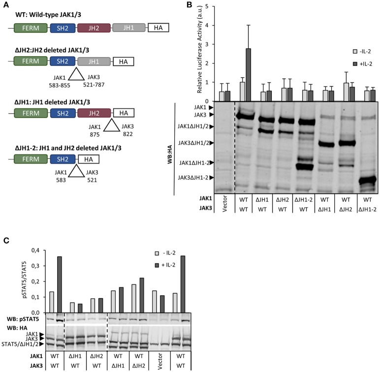
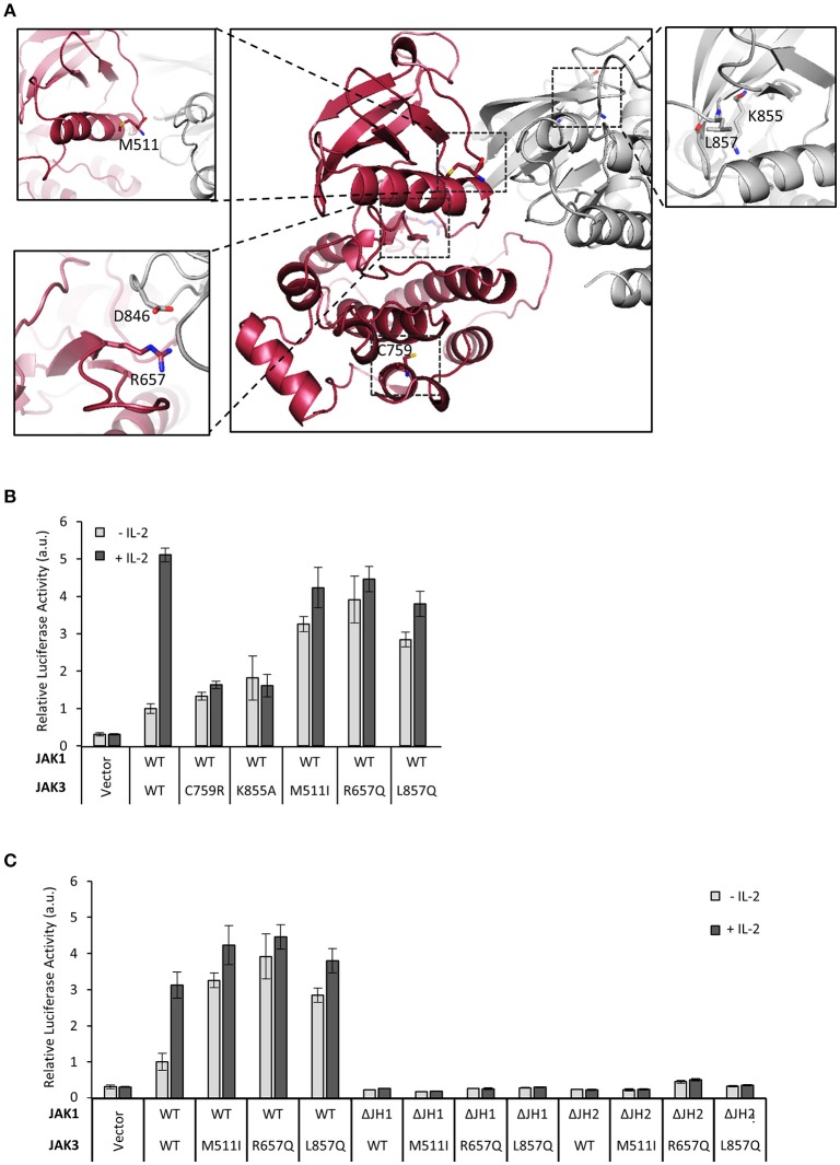
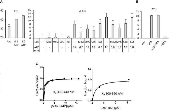
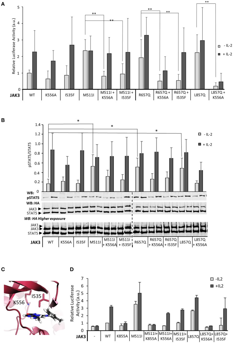
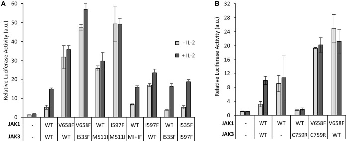
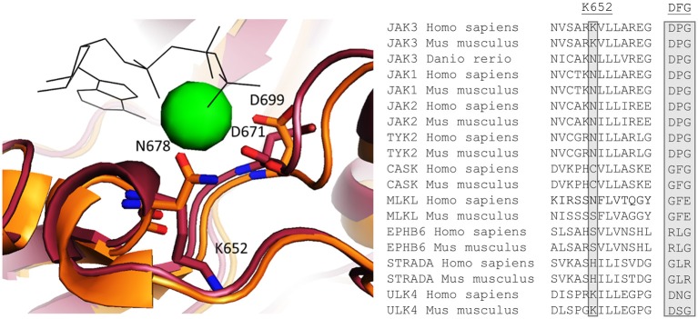
Janus kinase 3 (JAK3) tyrosine kinase has a central role in the control of lymphopoiesis, and mutations in JAK3 can lead to either severe combined immunodeficiency or leukemia and lymphomas. JAK3 associates with the common gamma chain (γc) receptor and functions in a heteromeric signaling pair with JAK1. In IL-2 signaling JAK1 is the effector kinase for STAT5 phosphorylation but the precise molecular regulatory mechanisms of JAK1 and JAK3 and their individual domains are not known. The pseudokinase domain (JAK homology 2, JH2) of JAK3 is of particular interest as approximately half of clinical JAK3 mutations cluster into it. In this study, we investigated the role of JH2s of JAK1 and JAK3 in IL-2R signaling and show that STAT5 activation requires both JH1 and JH2 of JAK1, while both JH1 and JH2 in JAK3 are specifically required for the cytokine-induction of cellular signaling. Characterization of recombinant JAK3 JH2 in thermal shift assay shows an unstable protein domain, which is strongly stabilized by ATP binding. Unexpectedly, nucleotide binding to JAK3 JH2 was found to be cation-independent. JAK3 JH2 showed higher nucleotide binding affinity in MANT-ATP and fluorescent polarization competition assays compared to the other JAK JH2s. Analysis of the functional role of ATP binding in JAK3 JH2 in cells and in zebrafish showed that disruption of ATP binding suppresses ligand-independent activation of clinical JAK3 gain-of-function mutations residing in either JH2 or JH1 but does not inhibit constitutive activation of oncogenic JAK1. ATP-binding site mutations in JAK3 JH2 do not, however, abrogate normal IL-2 signaling making them distinct from JH2 deletion or kinase-deficient JAK3. These findings underline the importance of JAK3 JH2 for cellular signaling in both ligand-dependent and in gain-of-function mutation-induced activation. Furthermore, they identify the JH2 ATP-binding site as a key regulatory region for oncogenic JAK3 signaling, and thus a potential target for therapeutic modulation.

Keywords: JAK kinase; cytokine; leukemia; nucleotide binding; pseudokinase.


Figures

Figure 1
Figure 1
Both JH1 and JH2 in JAK1 and JAK3 are required for functional IL-2 signaling. (A) Schematic presentation of JAK1 and JAK3 wild-type (WT) and domain deletion constructs. (B) Both JH1 and JH2 of JAK1 and JAK3 are required for IL-2 dependent STAT5 activation. U4Cγβ cells were transfected with JAK1 WT, JAK3 WT and domain deletion constructs together with STAT5 responsive reporter SPI-luc2 (28), either with Renilla or pRL-TK control vector (see Materials and Methods) and starved/stimulated. Similar results were obtained with both control vectors. Data shows averages and SD from normalized values combined from three independent experiments, each having triplicate samples. The expression of JAK1/JAK3 constructs was analyzed by anti-HA Western blotting. (C) IL-2-induced pSTAT5 requires both JH1 and JH2 in JAK1 and JAK3. STAT5 phosphorylation and expression of JAK1, JAK3 and STAT5 were detected from cell lysates from transfected U4Cγβ cells by immunoblotting with HA- and pTyr694 STAT5 antibodies. Similar results were obtained in three independent experiments.
Figure 2
Figure 2
Mutations in JAK3 JH2 can alter the function of the IL-2 pathway. (A) Localizations of the JAK3 mutants used in this article. Homology model of JAK3 JH2-JH1 (based on the TYK2 structure in Protein Data Bank (PDB) code: 4OLI) and the JAK3 JH1 structure [PDB code: 1YVJ (38)] were aligned with the JH1-JH2 structure of TYK2 (PDB code: 4OLI). JH2 is shown in red and JH1 in gray. Close-ups of the mutations are shown in subpanels. C759 is shown in the main picture. In the lower left panel, the (possible) interaction pair R657–D864 (in JH1) is shown. In the right panel, ATP was aligned from ATP-bound form of PKA [PDB code: 1ATP (39)]. (B) JAK3 JH2 C759R mutation efficiently inhibits IL-2 signaling and activating mutations in JAK3 JH2 and JH1. Luciferase assay of STAT5 activation in U4Cγβ cells transfected with JAK1 WT and JAK3 point mutations, SPI-luc2 reporter and β-galactosidase control. Values were normalized to JAK1 WT and JAK3 WT transfected sample Errors bars show SD of triplicate samples. Similar results were obtained using pRL-TK as a control. (C) Activating JAK3 mutants rely on both JH1 and JH2 of JAK1 for constitutive downstream signaling. SPI-luc2 reporter was used to detect the activation of STAT5 in U4Cγβ cells. Cells were transfected with WT or domain deletion JAK1 and WT or constitutively active JAK3 mutants. β-galactosidase plasmid was used as an internal control for the transfection efficiency. Values were normalized to WT JAK1 and JAK3. Error bars are SD of triplicate samples.
Figure 3
Figure 3
Nucleotide binding to recombinant JAK3 JH2. (A) JAK3 JH2 binds ATP without cations. The effect of ATP on Tm of JAK3 JH2 is shown in the left panel. The following graph shows changes in the melting temperature (ΔTm) in JAK3 JH2 in the presence of different cations and ATP. (B) Chelating agent does not affect ATP-induced increase on Tm of JAK3 JH2. DSF analysis performed in the presence of ATP (1 mM) and chelating agent EDTA (2 mM). (C) MANT-ATP binding assay with recombinant JAK3 JH2. Both protein and MANT-ATP were titrated and both experiments gave similar Kd in the sub-micromolar range. Left panel: titrating MANT-ATP to 1.5 μM JAK3-JH2. Kd range 330–440 nM. Right panel: titrating JAK3 JH2 to 0.25 μM MANT-ATP, Kd 500 nM. Protein concentrations were 0; 0.05; 0.25; 1.25 and 6.25 μM. In both figures, the Y-axis is the fraction of ligand-bound protein in relation to the total protein amount ([PL/Ptot]). The MANT-ATP titration was repeated three times and all experiments are plotted in the lower left panel. Protein titration was repeated twice and both experiments are blotted in the figure.
Figure 4
Figure 4
Inhibition of ATP binding to the pseudokinase domain of JAK3 reverts pathogenic, ligand-independent JAK3 signaling, while retaining cytokine-responsiveness. (A) JAK3 JH2 ATP-binding site mutations suppress activating JAK3 mutants. Luciferase assay was performed in U4Cγβ cells transfected with JAK1 and the indicated JAK3 mutants using β-galactosidase as a transfection control. The results are from three independent experiment, each having triplicate samples (n = 9, expect for K556A n = 7). Similar results were obtained with pRL-TK control. Data is normalized to the basal wild-type sample, errors are presented as SD. Statistical analysis of the activating mutations, and their corresponding double mutants were done with two-tailed t-test using unequal variances. **p < 0.001. (B) Representative Western blot of the transiently transfected U4Cγβ cell lysates showing the pSTAT5 signal. HA-tagged STAT5 was co-transfected with HA-tagged JAK1 and JAK3 and the lysates were blotted with anti-HA and anti-pSTAT5. Lower panel: Quantitative pSTAT5/STAT5 signal analysis. Activation of JAK3 signaling by M511I, R657Q and L857Q is significant (*p < 0.05). Data is average of five experiments. (C) The localization of the mutated JAK3 JH2 ATP-binding residues. Homology model of JAK3 JH2 (based on TYK2 structure from PDB, code: 4OLI), ATP is shown in gray. (D) Kinase-inactive JAK3 K855A abolishes IL-2 signaling in the presence of the activating JAK3 mutation M511I. Luciferase assay with STAT5-responsive promoter and with pRL-TK control.
Figure 5
Figure 5
Disrupting the JAK3 JH2 nucleotide binding site inhibits JAK3-induced, but not JAK1-induced, hyperactivation, and vice versa. (A) Constitutive activation of STAT5 caused by oncogenic JAK3 mutations are suppressed only by ATP-binding site mutations in JAK3 and not its dimerization partner JAK1. Luciferase assay of U4Cγβ cells transiently transfected with JAK1, JAK3, SPI-luc2 and pRL-TK. Results are average from two separate experiments each with triplicate or duplicate (I597F+I535F and I597F+WT JAK3) samples. The results are normalized to the JAK1 WT and JAK3 WT sample. Error bars show the SD. (B) Inhibitory JAK3 SCID mutation C759R does not reduce oncogenic hyperaction of JAK1-mutant. Luciferase assay in U4Cγβ cells transiently transfected with JAK1, JAK3, SPI-luc2, and pRL-TK. Error bars are SD of triplicate samples and the data is representative of three independent experiments. Supplementary Figure 5B shows immunoblotting for protein expression levels.
Figure 6
Figure 6
Left: Homology model of JAK3 JH2 aligned with the crystal structure of JAK2 [PDB code: 4FVQ (25)]. JAK3 is shown in red and JAK2 in orange. ATP is shown as black shits and the Mg2+ present in the JAK2 JH2 structure as a green sphere. Right: An alignment of human and mice sequences of JAKs and several other pseudokinases for Mg2+ coordination, JAK3 K652, which is conserved as N in other JAKs, and is important for Mg2+ coordination, is highlighted. Also the canonical DFG motif are compared and show variation between the pseudokinases.

References

    1. Yamaoka K, Saharinen P, Pesu M, Holt VET, Silvennoinen O, O'Shea JJ. The Janus kinases (Jaks). Genome Biol. (2004) 5:253. 10.1186/gb-2004-5-12-253 - DOI - PMC - PubMed
    1. Ghoreschi K, Laurence A, O'Shea JJ. Janus kinases in immune cell signaling. Immunol Rev. (2009) 228:273–87. 10.1111/j.1600-065X.2008.00754.x - DOI - PMC - PubMed
    1. Degryse S, de Bock CE, Cox L, Demeyer S, Gielen O, Mentens N, et al. . JAK3 mutants transform hematopoietic cells through JAK1 activation, causing T-cell acute lymphoblastic leukemia in a mouse model. Blood (2014) 124:3092–100. 10.1182/blood-2014-04-566687 - DOI - PubMed
    1. Walters DK, Mercher T, Gu TL, O'Hare T, Tyner JW, Loriaux M, et al. . Activating alleles of JAK3 in acute megakaryoblastic leukemia. Cancer Cell (2006) 10:65–75. 10.1016/j.ccr.2006.06.002 - DOI - PubMed
    1. Malinge S, Ragu C, Della-Valle V, Pisani D, Constantinescu S, Perez C, et al. . Activating mutations in human acute megakaryoblastic leukemia. Blood (2008) 112:4220–6. 10.1182/blood-2008-01-136366 - DOI - PubMed

LinkOut - more resources