. 2019 Jan;25(1):73-81.
doi: 10.3201/eid2501.180807.

Variable Protease-Sensitive Prionopathy Transmission to Bank Voles

Variable Protease-Sensitive Prionopathy Transmission to Bank Voles

Romolo Nonno et al. Emerg Infect Dis. 2019 Jan.

Abstract

Variably protease-sensitive prionopathy (VPSPr), a recently described human sporadic prion disease, features a protease-resistant, disease-related prion protein (resPrPD) displaying 5 fragments reminiscent of Gerstmann-Sträussler-Scheinker disease. Experimental VPSPr transmission to human PrP-expressing transgenic mice, although replication of the VPSPr resPrPD profile succeeded, has been incomplete because of second passage failure. We bioassayed VPSPr in bank voles, which are susceptible to human prion strains. Transmission was complete; first-passage attack rates were 5%-35%, and second-passage rates reached 100% and survival times were 50% shorter. We observed 3 distinct phenotypes and resPrPD profiles; 2 imitated sporadic Creutzfeldt-Jakob disease resPrPD, and 1 resembled Gerstmann-Sträussler-Scheinker disease resPrPD. The first 2 phenotypes may be related to the presence of minor PrPD components in VPSPr. Full VPSPr transmission confirms permissiveness of bank voles to human prions and suggests that bank vole PrP may efficiently reveal an underrepresented native strain but does not replicate the complex VPSPr PrPD profile.

Keywords: 109I; 109M; 7 kDa; Creutzfeldt-Jakob; Gerstmann-Sträussler-Scheinker; VPSPr; Variable protease-sensitive prionopathy; atypical glycoform; bank voles; lesion profile; minor PrP components; phenotype; prion disease; prions; sporadic; strain; substrain; transmissibility; type 1; type 2.


Figures
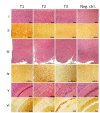
Figure 1
Figure 1
Histopathology and prion protein (PrP) immunohistochemistry images of brain regions from variably protease-sensitive prionopathy (VPSPr)–inoculated bank voles 109I harboring the histopathologic phenotypes T1, T2, or T3. For T1 bank voles, the cerebral neocortex (i, ii) shows moderate spongiform degeneration with substantial PrP immunostaining often displaying a laminar enhancement (bracket), but spongiform degeneration and PrP immunostaining were minimal or lacking in T2 and T3 bank voles (i, ii, T2, T3). By contrast, spongiform degeneration and immunostaining characterized by granular aggregates were selectively prominent in the hypothalamus of T2 but not in T1 and T3 (iii, iv) bank voles. Distinct lesions also occurred in a region comprising dorsal hippocampus and overlying hemispheric white matter (v, vi) where T1 shows hippocampal spongiform degeneration with fine disease-related prion protein (PrPD) deposition; T2 shows granular or small plaque-like PrPD deposition affecting primarily the alveus; and T3 shows an intense and compact PrPD deposition affecting stratum oriens, alveus, and white matter of the corpus callosum. The negative control (Neg Ctrl) column (i–vi) shows tissue from inoculated bank voles that were negative by Western blot. Neither spongiform degeneration nor positive PrP immunostaining was observed in these controls. All panels T1–T3 are from second passage; antibody SAF84. Scale bars for row iii indicate 500 μm; all others indicate 100 μm. This figure is also available online at https://wwwnc.cdc.gov/EID/article/25/1/18-0807-F1.htm.
Figure 2
Figure 2
Profiles of topographic distribution and severity of spongiform degeneration in the brains of bank voles harboring T1–T3 phenotypes after inoculation with brain homogenate from variably protease sensitive prionopathy (VPSPr) and control bank voles inoculated with sCJD. A) Spongiform degeneration characterized both T1 and T2 phenotypes but displayed significantly divergent distributions in 5 of the 10 anatomic locations examined; spongiform degeneration affected primarily the cerebral cortex in T1 and the hypothalamus and brain stem in T2; no difference in vacuolar mean diameter was observed between T1 and T2. Spongiform degeneration scores associated with the T3 phenotype were minimal or absent in most locations except for the white matter, especially in the corpus callosum, which was virtually unaffected in T1 and T2. *p<0.5; **p<0.006; ***p<0.0001 of T1 versus T2 and T3 WM versus T1 and T2; inocula: T1 and T2 VPSPr-129MV, T3 VPSPr-129MM; vacuoles measured (n = ≈2,000) in T1 and T2 combined. B) Comparative study of T1 profiles generated in bv109M and bv109I revealed an overall more severe spongiform degeneration in bv109I but no significant difference in distribution (†, p<0.001, ‡, p<0.003; N = 3 Bv109I and Bv109M). The T1 spongiform degeneration profile generated by bv109M after inoculation with VPSPr-129MM reproduced the profile generated with sCJDMM1 extracts used as control for human type 1 (bv109M N = 3 for each profile). Similar results were obtained when comparing the T1 profile of bv109I inoculated with VPSPr-129MV and profiles of bv109I inoculated with sCJDMV1 (data not shown). BN, basal nuclei; BSs and BSi, brainstem superior and inferior; bv, bank vole; CC, cerebral cortex; Ce, cerebellum; ctrl, control; Hi, hippocampus; Hth, hypothalamus; sCJD, sporadic Creutzfeldt-Jakob disease; Sept.N, septal nuclei; Th, thalamus; WM, white matter.
Figure 3
Figure 3
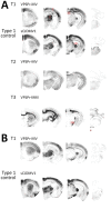
Representative paraffin-embedded tissue (PET) blots of protease-resistant, disease-related prion protein (resPrPD) distribution in phenotypes T1–T3 and controls. A) For T1, PrPD predominated in cerebral cortex (C), thalamus (T), superior colliculus (SC), lateral geniculate nucleus (LG), and substantia nigra (SN). A similar PrPD distribution was observed with transmission of sCJDMV1 used as type control. T2 showed a more uniform distribution in cerebral cortex and subcortical nuclei of an apparently lesser amount of PrPD; T3 appeared to preferentially affect the hemispheric white matter and other subcortical regions such as the alveus, the corpus callosum, the anterior commissure, and fascicles surrounding thalamus as well as other white matter formations such as fimbria, brachium of superior colliculus, medial lemniscus, and cerebral peduncles. Small amounts of PrPD were also observed in cerebellar and medullary white matter (asterisk [*]). B) PrPD T1 distribution resembled that of bank voles (bv) 109I after transmission of the same VPSPr-MV brain homogenate (compare with T1 in A). A similar distribution was also observed after inoculation with sCJDMV1. Left to right: coronal sections of telencephalon midlevel caudate nucleus; diencephalon midlevel thalamus; midbrain; and hindbrain-level medulla and cerebellum. sCJD, sporadic Creutzfeldt-Jakob disease; VPSPr, variably protease-sensitive prionopathy.
Figure 4
Figure 4
Immunoblot characteristics of protease-resistant, disease-related prion protein (resPrPD) distribution in phenotypes T1–T3 and controls. Regular and long exposures revealed the overall similarity of the 3-band profiles in T1 and T2, but resPrPD profile, including glycoform representation, differed in the 2 phenotypes with all 3 monoclonal antibodies (Ab) used. T1 included a 7-kDa band, not detected in T2, similar to mobility and Ab immunoreactivity of the T3 7-kDa fragment. The T1 profile matched the profile generated in isogenic bank voles inoculated with sCJDMV1 used as human resPrPD type 1 control (ctrl) (lane 2). The T3 profile, visible only after long film exposures, featured a 7-kDa band, but slower migrating bands with variable immunoreactivity were also visible. None of the T1–T3 profiles matched the original VPSPr profile (first lane) although the ≈7-kDa and both 23-kDa and 19-kDa bands were shared with T1 and T2, respectively (compare first with T1 and T2 lanes). The complexity of the native resPrPD profile from VPSPr homogenate is demonstrated by probing with 1E4, a monoclonal Ab to human PrP highly reactive to VPSPr resPrPD (top right panel) (6). Monoclonal Ab 12B2 (middle panels) with high affinity for human resPrPD type 1 confirmed the type 1 characteristics of the resPrPD associated with the T1 phenotype. The small amount of resPrPD type 1 in 1 T2 bank vole probably represents incomplete proteinase K (PK) digestion (lane 5, right panel) (19). Monoclonal Ab SAF84 to the PrP C-terminus, unreactive to human PrP, further underlined the divergence in resPrPD primary structure in T1 and T2 compared with T3. Aside from revealing an additional 13-kDa fragment, strongly detected in T1 and T2 and weakly in T3, SAF84 did not detect the 7-kDa fragment, supporting its internal origin (i.e., cleaved at both N- and C-termini). Uninoculated bank voles were negative for resPrPD. All samples were PK treated. sCJD, sporadic Creutzfeldt-Jakob disease; uninoc., not inoculated; polym., polymorphism; VPSPr, variably protease-sensitive prionopathy.
Figure 5
Figure 5
Glycoform ratio of protease-resistant, disease-related prion protein (resPrPD) in phenotypes T1 and T2. The ratio of resPrPD associated with T1 (T1 109I) was 48% for diglycosylated, 44% for monoglycosylated, and 8% for unglycosylated conformers and significantly differed in each glycoform from the 17%, 63%, and 20% corresponding ratio of T2 (T2 109I). *p<0.0001; †p<0.005; ‡p<0.05. Glycoform ratios of T1 109I and T1 109M as well as that of type 1 control (from bank voles 109I inoculated with sporadic Creutzfeldt-Jakob disease MV1 and used as human type 1 controls) did not significantly differ from each other. Each bar represents mean ± SD of n = 4 for T1 109M, n = 6 for T1 109I, n = 6 for T2, and n = 2 for type 1 control.
Figure 6
Figure 6
Relative quantities of totPrPD and resPrPD in T1–T3 phenotypes. totPrPD accounted for 93.1% and resPrPD for 81.3% of total PrP recovered from bank voles harboring the T1 phenotype. Corresponding percentages for T2 were 91.0% and 33.0%, and T3 totPrPD and resPrPD accounted only for 8.0% and 0.2% of total PrP and differed significantly from both T1 and T2 in each of the 2 components. ResPrPD also differed significantly between T1 and T2 (each bar represents the mean ± SD of n = 3 T1, n = 3 T2, and n = 5 T3; all data are from bank voles 109I; antibody 9A2). bv, bank vole; resPrPD protease-resistant, disease-related prion protein; totPrPD, comprising protease-sensitive PrPD and resPrPD. *p<0.0001; †p<0.0001 vs. T1 and p<0.05 vs. T2; ‡p<0.01.

References

    1. Gambetti P. Creationism and evolutionism in prions. Am J Pathol. 2013;182:623–7. 10.1016/j.ajpath.2012.12.016 - DOI - PMC - PubMed
    1. Gambetti P, Cali I, Notari S, Kong Q, Zou WQ, Surewicz WK. Molecular biology and pathology of prion strains in sporadic human prion diseases. Acta Neuropathol. 2011;121:79–90. 10.1007/s00401-010-0761-3 - DOI - PMC - PubMed
    1. Puoti G, Bizzi A, Forloni G, Safar JG, Tagliavini F, Gambetti P. Sporadic human prion diseases: molecular insights and diagnosis. Lancet Neurol. 2012;11:618–28. 10.1016/S1474-4422(12)70063-7 - DOI - PubMed
    1. Parchi P, Strammiello R, Giese A, Kretzschmar H. Phenotypic variability of sporadic human prion disease and its molecular basis: past, present, and future. Acta Neuropathol. 2011;121:91–112. 10.1007/s00401-010-0779-6 - DOI - PubMed
    1. Poggiolini I, Saverioni D, Parchi P. Prion protein misfolding, strains, and neurotoxicity: an update from studies on mammalian prions. Int J Cell Biol. 2013;2013:910314. - PMC - PubMed

Publication types