

DukeSim: A Realistic, Rapid, and Scanner-Specific Simulation Framework in Computed Tomography
- PMID: 30561344
- PMCID: PMC6598436
- DOI: 10.1109/TMI.2018.2886530
DukeSim: A Realistic, Rapid, and Scanner-Specific Simulation Framework in Computed Tomography
Abstract
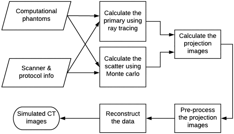
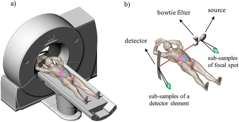
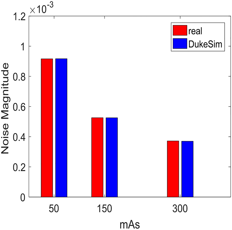
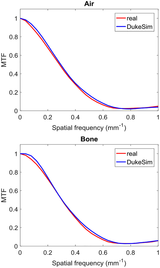
The purpose of this study was to develop a CT simulation platform that is: 1) compatible with voxel-based computational phantoms; 2) capable of modeling the geometry and physics of commercial CT scanners; and 3) computationally efficient. Such a simulation platform is designed to enable the virtual evaluation and optimization of CT protocols and parameters for achieving a targeted image quality while reducing radiation dose. Given a voxelized computational phantom and a parameter file describing the desired scanner and protocol, the developed platform DukeSim calculates projection images using a combination of ray-tracing and Monte Carlo techniques. DukeSim includes detailed models for the detector quantum efficiency, quantum and electronic noise, detector crosstalk, subsampling of the detector and focal spot areas, focal spot wobbling, and the bowtie filter. DukeSim was accelerated using GPU computing. The platform was validated using physical and computational versions of a phantom (Mercury phantom). Clinical and simulated CT scans of the phantom were acquired at multiple dose levels using a commercial CT scanner (Somatom Definition Flash; Siemens Healthcare). The real and simulated images were compared in terms of image contrast, noise magnitude, noise texture, and spatial resolution. The relative error between the clinical and simulated images was less than 1.4%, 0.5%, 2.6%, and 3%, for image contrast, noise magnitude, noise texture, and spatial resolution, respectively, demonstrating the high realism of DukeSim. The runtime, dependent on the imaging task and the hardware, was approximately 2-3 minutes per rotation in our study using a computer with 4 GPUs. DukeSim, when combined with realistic human phantoms, provides the necessary toolset with which to perform large-scale and realistic virtual clinical trials in a patient and scanner-specific manner.
Figures


References
-
- Tsui B et al. , “Quantitative cardiac SPECT reconstruction with reduced image degradation due to patient anatomy,” IEEE transactions on nuclear science, vol. 41, no. 6, pp. 2838–2844, 1994.
-
- Lee C, Williams JL, Lee C, and Bolch WE, “The UF series of tomographic computational phantoms of pediatric patients,” Medical physics, vol. 32, no. 12, pp. 3537–3548, 2005. - PubMed
-
- Zhang SX, Heng PA, and Liu ZJ, “Chinese visible human project,” Clinical Anatomy, vol. 19, no. 3, pp. 204–215, 2006. - PubMed
-
- Cassola V, de Melo Lima V, Kramer R, and Khoury H, “FASH and MASH: female and male adult human phantoms based on polygon mesh surfaces: I. Development of the anatomy,” Physics in medicine and biology, vol. 55, no. 1, p. 133, 2009. - PubMed
