. 2019 Sep 27;69(8):1345-1352.
doi: 10.1093/cid/ciy1066.

Human Immunodeficiency Virus Type 1 RNA Detected in the Central Nervous System (CNS) After Years of Suppressive Antiretroviral Therapy Can Originate from a Replicating CNS Reservoir or Clonally Expanded Cells

Affiliations

Human Immunodeficiency Virus Type 1 RNA Detected in the Central Nervous System (CNS) After Years of Suppressive Antiretroviral Therapy Can Originate from a Replicating CNS Reservoir or Clonally Expanded Cells

Sarah B Joseph et al. Clin Infect Dis. .

Abstract

Background: Human immunodeficiency virus type 1 (HIV-1) populations are detected in cerebrospinal fluid (CSF) of some people on suppressive antiretroviral therapy (ART). Detailed analysis of these populations may reveal whether they are produced by central nervous system (CNS) reservoirs.

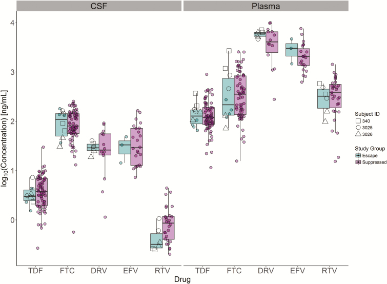
Methods: We performed a study of 101 asymptomatic participants on stable ART. HIV-1 RNA concentrations were cross-sectionally measured in CSF and plasma. In participants with CSF HIV-1 RNA concentrations sufficient for analysis, viral populations were genetically and phenotypically characterized over multiple time points.

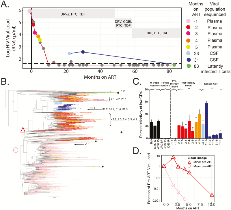
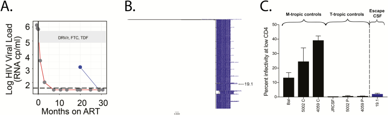
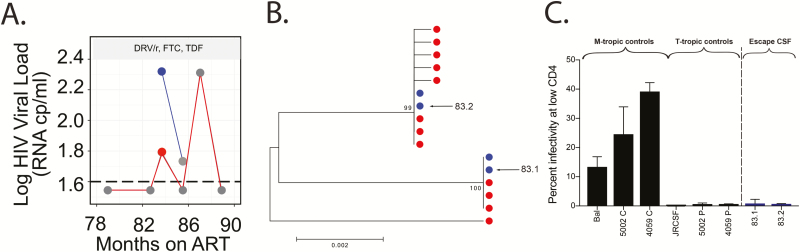
Results: For 6% of participants (6 of 101), the concentration of HIV-1 RNA in their CSF was ≥0.5 log copies/mL above that of plasma (ie, CSF escape). We generated viral envelope sequences from CSF of 3 participants. One had a persistent CSF escape population that was macrophage-tropic, partially drug resistant, genetically diverse, and closely related to a minor macrophage-tropic lineage present in the blood prior to viral suppression and enriched for after ART. Two participants (1 suppressed and 1 not) had transient CSF escape populations that were R5 T cell-tropic with little genetic diversity.

Conclusions: Extensive analysis of viral populations in 1 participant revealed that CSF escape was from a persistently replicating population, likely in macrophages/microglia, present in the CNS over 3 years of ART. CSF escape in 2 other participants was likely produced by trafficking and transient expansion of infected T cells in the CNS. Our results show that CNS reservoirs can persist during ART and that CSF escape is not exclusively produced by replicating CNS reservoirs.

Keywords: CNS; CSF escape; HIV reservoirs; drug resistance; persistence.


Figures

Figure 1.
Figure 1.
Persistent asymptomatic CSF escape variants are macrophage-tropic, produced by a population of infected cells in the central nervous system during ART, and genetically similar to macrophage-tropic variants detected in the plasma after ART initiation (participant 3026). A, HIV-1 viral loads in the plasma (values connected by a red line) and CSF (values connected by a blue line) were measured at multiple time points for participant 3026. HIV-1 was suppressed in the blood plasma after 10 months of ART, and CSF escape was observed after 23 and 31 months of ART but undetectable after 83 months of ART. Nongray dots indicate time points at which viral RNA was extracted from plasma and/or CSF and a MiSeq with Primer ID approach was used to generate partial env sequences or an outgrowth analysis was performed. ART regimens are shown in gray boxes. B, A neighbor-joining phylogenetic tree was constructed to compare partial env sequences from the plasma and CSF at multiple time points. Prior to ART, 92% of virus in the plasma formed a single major lineage (node marked with a pink circle), 4% of the plasma population was found in a separate, minor lineage (node marked with a red triangle), and the remaining virus was found in 4 small lineages representing approximately 4% of the pre-ART plasma population. After 2 months of ART, the initially rare lineage represented 82% of variants in the plasma. CSF escape variants were largely monophyletic and most closely related to the lineage that was rare pre-ART. Single-genome amplification was used to amplify 15 full-length env genes from the CSF and plasma at multiple time points (amplicon names indicate months on ART and are designated on the tree) and amplicons were cloned into expression vectors for entry phenotype analyses. C, Cloned HIV-1 envs were used to produce Env-pseudotyped reporter viruses and perform single-cycle infection of CD4lowCCR5high Affinofile cells to determine whether clones were macrophage-tropic. The entry phenotypes of previously characterized macrophage-tropic and T cell-tropic controls are shown. Two clones from the blood population pre-ART, one from the majority lineage pre-ART (pink circle) and another from elsewhere in the tree, were found to be T cell-tropic due to their inability to efficiently enter cells expressing a low density of CD4. In contrast, envs cloned from the initially rare lineage (red triangle) and CSF escape variants were all macrophage-tropic. D, Sequence and viral load data were used to estimate the viral load of the lineages at each time point and to plot the viral load of each lineage as a fraction of its pre-ART viral load. Prior to ART (to the left of the vertical line), each lineage starts at 1, but by 5 months post-ART, the initially common lineage (pink circle) had decayed 3 log10 from its pre-ART frequency, while the initially rare lineage (red triangle) only decayed 0.7 log10. This difference was not simply due to differences in the post-ART decay rate but also due to expansion of this population near the time of ART initiation. Abbreviations: ART, antiretroviral therapy; BIC, bictegravir; COBI, cobicistat; CSF, cerebrospinal fluid; DRV/r, ritonavir-boosted darunavir; FTC, emtricitabine; HIV, human immunodeficiency virus; TAF, tenofovir alafenamide; TDF, tenofovir disoproxil fumarate.
Figure 2.
Figure 2.
Episodic asymptomatic CSF escape variants are most likely produced by clonally expanded CD4+ T cells present in the CNS during ART (participant 3025). A, HIV-1 viral loads in the plasma (values connected by a red line) and CSF (values connected by a blue line) were measured at multiple time points for participant 3025. CSF escape was observed after 19 months of ART but was undetectable at 27 months post-ART. A MiSeq with Primer ID approach was used to generate partial env sequences from the CSF escape population (blue dot). ART regimen is shown in the gray box. B, A neighbor-joining phylogenetic tree of 733 partial env sequences illustrates that the CSF escape population was nearly homogeneous. C, A full-length HIV-1 env cloned from this homogeneous population was pseudotyped and shown to be T cell-tropic in a CD4lowCCR5high Affinofile entry assay. Abbreviations: ART, antiretroviral therapy; CSF, cerebrospinal fluid; DRV/r, ritonavir-boosted darunavir; FTC, emtricitabine; HIV, human immunodeficiency virus; TDF, tenofovir disoproxil fumarate.
Figure 3.
Figure 3.
The source of asymptomatic CSF escape in a poorly suppressed participant is unclear (participant 340). A, HIV-1 viral loads in the plasma (values connected by a red line) and CSF (values connected by a blue line) were measured at multiple time points for participant 340. Nongray dots indicate the time point at which viral RNA was extracted from plasma and/or CSF, and single-genome amplification (SGA) was used to generate full-length env genes. Antiretroviral therapy regimen is shown in the gray box. B, A neighbor-joining phylogenetic tree of full-length env sequences illustrates that identical sequences are found in the CSF and blood plasma. C, Full-length HIV-1 envs cloned from the CSF population were pseudotyped and shown to be T cell-tropic based on an inability to efficiently enter Affinofile cells expressing a low surface density of CD4. Abbreviations: ART, antiretroviral therapy; CSF, cerebrospinal fluid; DRV/r, ritonavir-boosted darunavir; FTC, emtricitabine; HIV, human immunodeficiency virus; TDF, tenofovir disoproxil fumarate.
Figure 4.
Figure 4.
Individuals with CSF escape had drug concentrations similar to those of well-suppressed individuals. CSF and plasma collected from the THINC (Tropism of HIV-1, Inflammation and NeuroCognition) cohort were analyzed for the concentration of 5 of the most common drugs used in the cohort. As expected, CSF drug concentrations were lower than that of plasma. On average, drug concentrations were not lower in the individuals with CSF escape (green) relative to individuals who were virologically suppressed (purple). Abbreviations: CSF, cerebrospinal fluid; DRV, darunavir; EFV, efavirenz; FTC, emtricitabine; RTV, ritonavir; TDF, tenofovir disoproxil fumarate.

References

    1. Dinoso JB, Kim SY, Wiegand AM, et al. . Treatment intensification does not reduce residual HIV-1 viremia in patients on highly active antiretroviral therapy. Proc Natl Acad Sci U S A 2009; 106:9403–8. - PMC - PubMed
    1. Doyle T, Smith C, Vitiello P, et al. . Plasma HIV-1 RNA detection below 50 copies/ml and risk of virologic rebound in patients receiving highly active antiretroviral therapy. Clin Infect Dis 2012; 54:724–32. - PubMed
    1. Maldarelli F, Palmer S, King MS, et al. . ART suppresses plasma HIV-1 RNA to a stable set point predicted by pretherapy viremia. PLoS Pathog 2007; 3:e46. - PMC - PubMed
    1. Hermankova M, Ray SC, Ruff C, et al. . HIV-1 drug resistance profiles in children and adults with viral load of <50 copies/ml receiving combination therapy. JAMA 2001; 286:196–207. - PubMed
    1. Kieffer TL, Finucane MM, Nettles RE, et al. . Genotypic analysis of HIV-1 drug resistance at the limit of detection: virus production without evolution in treated adults with undetectable HIV loads. J Infect Dis 2004; 189:1452–65. - PubMed

Publication types

MeSH terms