. 2019 Jul 24;141(29):11465-11470.
doi: 10.1021/jacs.8b12203. Epub 2019 Jan 2.

Atomic Structure of a Fluorescent Ag8 Cluster Templated by a Multistranded DNA Scaffold

Affiliations

Atomic Structure of a Fluorescent Ag8 Cluster Templated by a Multistranded DNA Scaffold

Dustin J E Huard et al. J Am Chem Soc. .

Abstract

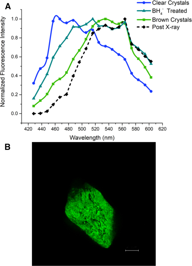
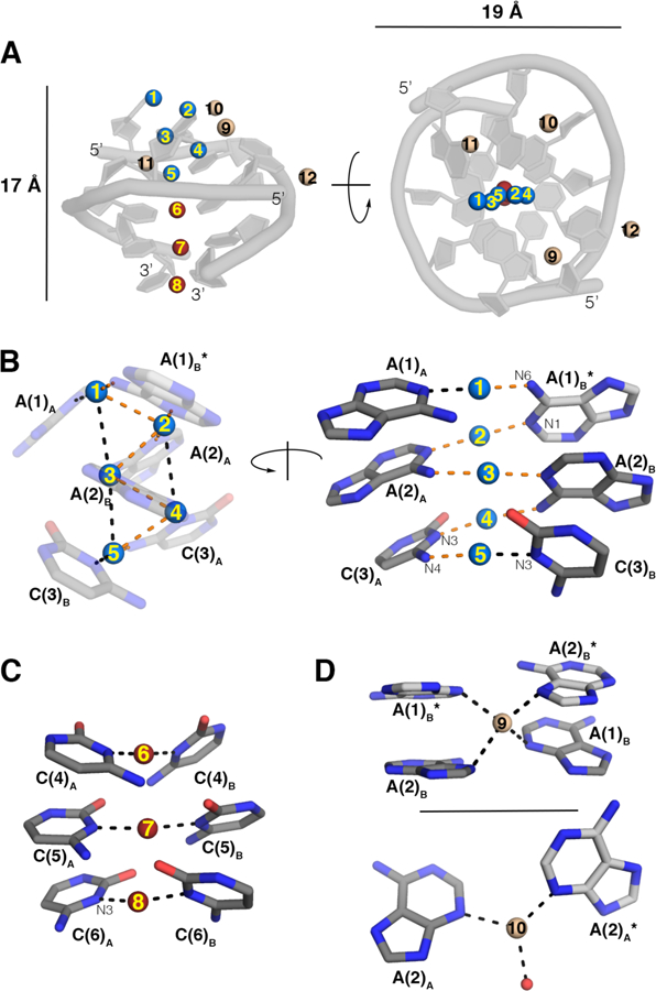
Multinuclear silver clusters encapsulated by DNA exhibit size-tunable emission spectra and rich photophysics, but their atomic organization is poorly understood. Herein, we describe the structure of one such hybrid chromophore, a green-emitting Ag8 cluster arranged in a Big Dipper-shape bound to the oligonucleotide A2C4. Three 3' cytosine metallo-base pairs stabilize a parallel A-form-like duplex with a 5' adenine-rich pocket, which binds a metallic, trapezoidal-shaped Ag5 moiety via Ag-N bonds to endo- and exocyclic nitrogens of cytosine and adenine. The unique DNA configuration, constrained coordination environment, and templated Ag8 cluster arrangement highlight the reciprocity between the silvers and DNA in adopting this structure. These first atomic details of a DNA-encapsulated Ag cluster fluorophore illuminate many aspects of biological assembly, nanoscience, and metal cluster photophysics.


Figures

Fig. 1
Fig. 1. Fluorescence from Ag:DNA crystals.
(A) Fluorescence spectra from Ag:DNA crystals excited at 405 nm. Emission shifts from blue to green upon chemical (BH4-) or x-ray-induced photoreduction, both of which resemble emission from initially brown crystals. Green emission from brown/reduced clusters is ~100 fold brighter than is the blue emission from non-reduced, clear, colorless crystals. (B) Central confocal optical section demonstrating green fluorescence from a chemically reduced crystal excited at 405nm. Scalebar 25 µm.
Fig. 2
Fig. 2. Arrangements of silvers.
(A). Two views of the A2C4 dimer with silvers and dimensions labeled. (B). Two zoomed views of the pentameric core. Left, Ag-Ag distances with orange dashes are ~2.9 Å; black dashes ~3.1–3.4 Å. Right, bidentate base-Ag interactions highlighted in orange are the same distance as monodentate interactions (black), ~2.1 Å. Subscripted letters denote DNA chain A or B, asterisks indicate symmetry related nucleobases. Representative coordinating nitrogens are labeled. (C). Zoomed-in view of near linear-coordinating silvers with 3’ cytosines. (D). Expanded view of silvers occupying special positions in the crystal (occupancy = 0.5), which coordinate adenines from symmetry-related molecules. Red sphere in D is ordered solvent. For C, D, black dashes are ~2.1 Å and the labeling scheme is as in (B). See also Tables S3 and S4, Fig. S3.
Fig. 3
Fig. 3. Configuration of A2C4 DNA strands in the asymmetric unit.
(A). Two views of the dimer in the asymmetric unit as in Fig. 2. Yellow, adenines. Purple, cytosines. (B). Zoomed-in view of hydrogen bonds (green dashes) in the stem region. Labels are as in Fig. 2B. (C). Superposition of chains A, B in the asymmetric unit, r.m.s.d. = 1.6 Å. See also Fig. S4, Tables S5 and S6.
Fig. 4
Fig. 4. Crystallographic metallotetraplex.
(A). Adenines from two clusters are in close proximity due to crystal packing and are stabilized by silvers on special positions in the crystal (see also Fig. 2D). Silvers are depicted as in Fig. 2 and colored as in Fig. 3. (B). Enlarged view showing central solvent molecule (orange) and ~3 Å hydrogen bonding distances of silvers (black dashes).

References

    1. Kubo R, Electronic properties of metallic fine particles. J. Phys. Soc. Jpn 1962, 17 (6), 975–986.
    1. Jin R, Atomically precise metal nanoclusters: stable sizes and optical properties. Nanoscale 2015, 7 (5), 1549–1565. - PubMed
    1. Peyser LA; Vinson AE; Bartko AP; Dickson RM, Photoactivated fluorescence from individual silver nanoclusters. Science 2001, 291 (5501), 103–106. - PubMed
    1. de Heer WA, The physics of simple metal clusters: experimental aspects and simple models. Rev. Mod. Phys 1993, 65 (3), 611–676.
    1. Zheng J; Nicovich PR; Dickson RM, Highly fluorescent noble-metal quantum dots. Annu. Rev. Phys. Chem 2007, 58, 409. - PMC - PubMed

Publication types