. 2018 Dec 17;10(12):720.
doi: 10.3390/v10120720.

A Divergent Hepatitis D-Like Agent in Birds

Affiliations

A Divergent Hepatitis D-Like Agent in Birds

Michelle Wille et al. Viruses. .

Abstract

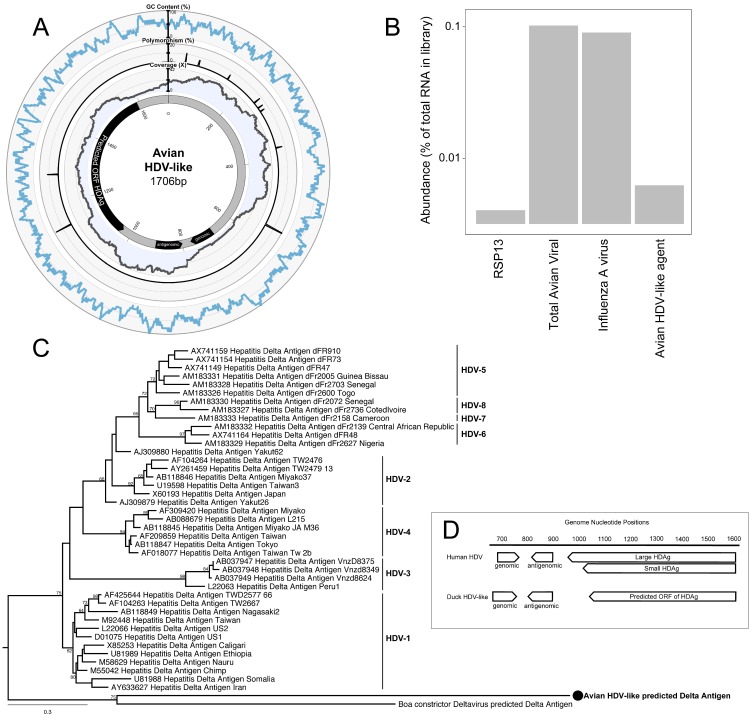
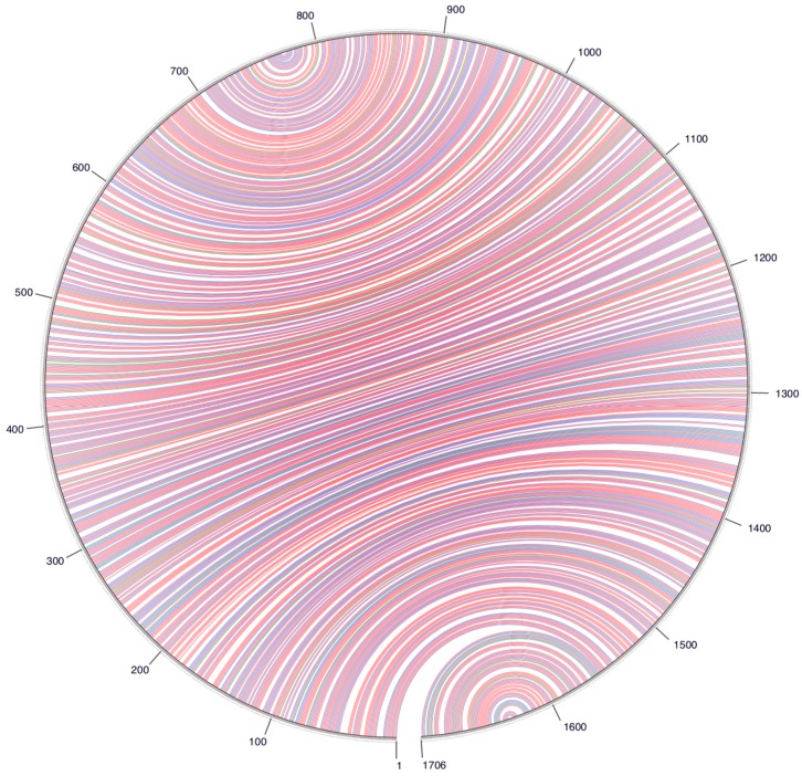
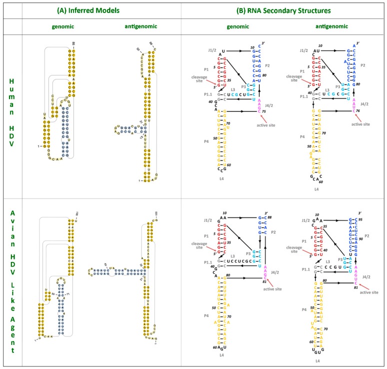
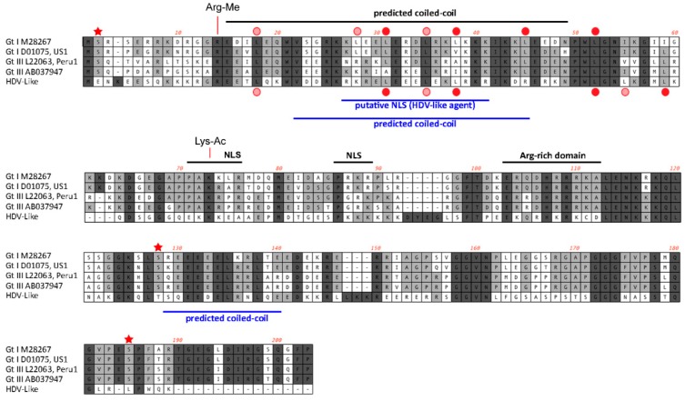
Hepatitis delta virus (HDV) is currently only found in humans and is a satellite virus that depends on hepatitis B virus (HBV) envelope proteins for assembly, release, and entry. Using meta-transcriptomics, we identified the genome of a novel HDV-like agent in ducks. Sequence analysis revealed secondary structures that were shared with HDV, including self-complementarity and ribozyme features. The predicted viral protein shares 32% amino acid similarity to the small delta antigen of HDV and comprises a divergent phylogenetic lineage. The discovery of an avian HDV-like agent has important implications for the understanding of the origins of HDV and sub-viral agents.

Keywords: co-evolution; dabbling duck; hepatitis D virus; phylogeny.


Conflict of interest statement

The authors declare no conflict of interest.

Figures

Figure 1
Figure 1
Characteristics of the genome of an avian hepatitis delta virus (HDV)-like agent. (A) Avian HDV-like agent genome, annotated with ORFs, genomic, and antigenomic ribozyme sites. Metadata rings include the read coverage, proportion of polymorphisms in reads, followed by GC content. (B) Abundance of transcripts in the metatranscriptomic library. Total avian viral abundance was dominated by that of the influenza A virus. However, the abundance of HDV is higher than that of Ribosomal protein S13 (RPS13), a stably expressed reference gene in Mallards (Anas platyrhynchos). (C) Maximum likelihood phylogeny of the HDAg protein. Representative human HDAg sequences fall into the currently described clades HDV1-8 (5). The scale bar represents the number of amino acid substitutions per site. The phylogeny is rooted between the human and avian/snake viruses. (D) Location of genomic and antigenomic ribozyme sequences, and the predicted ORF of the delta antigen in the avian HDV-like genome compared to their location in the HDV genome sequence (GenBank accession X04451.1).
Figure 2
Figure 2
A circle graph showing the base pairing of the circular RNA genome structure of the avian HDV-like agent into an unbranched rod-like structure. The circle circumference represents the genome sequence, and the arcs represent the base pairing. Colouring of arcs: Red for G-C pairing, blue for A-U pairing, green for G-U pairing, and yellow for other types of pairings.
Figure 3
Figure 3
HDV ribozymes. (A) Secondary structures of the genomic and antigenomic ribozymes inferred using the TT2NE algorithm. The HDV ribozyme models were used as reference to screen for the ribozyme sequences in the avian HDV-like genome sequence. (B) Re-drawn secondary structures of the genomic and antigenomic ribozymes based on the secondary structures shown in the review by Webb and Luptak (21).
Figure 4
Figure 4
Features of the predicted HDAg protein. Alignment of the amino acid sequences (small delta antigen) translated from the genomes of HDV and the avian HDV delta-like agent. The potential coiled-coil region is highlighted, including the presence of leucine residues in the correct spacing for a leucine zipper (filled red circle). The delta antigen does not have a strict requirement for leucine in the d-position of the heptad repeat. Additional leucine residues are shown by circles in light red. Serine residues that are conserved between different HDV genotypes and post-translationally modified (phosphorylated) are highlighted with an asterisk. The conserved arginine and lysine residues modified by methylation (Arg-Me) and acetylation (Lys-Ac) are indicated. NLS: Nuclear localisation signal.

References

    1. Taylor J., Pelchat M. Origin of hepatitis Delta virus. Future Microbiol. 2010;5:393–402. doi: 10.2217/fmb.10.15. - DOI - PMC - PubMed
    1. Taylor J.M., Purcell R.H., Farci P. Hepatitis D (D) virus. In: Knipe D.M., Howley P.M., editors. Fields Virology. Lippincott Williams & Wilkins; Philadelphia, PA, USA: 2013. pp. 2222–2241.
    1. Wedemeyer H., Manns M.P. Epidemiology, pathogenesis and management of hepatitis D: Update and challenges ahead. Nat. Rev. Gastroenterol. Hepatol. 2010;7:31–40. doi: 10.1038/nrgastro.2009.205. - DOI - PubMed
    1. WHO Hepatitis D Fact Sheet. [(accessed on 24 February 2018)];2017 Available online: http://www.who.int/mediacentre/factsheets/hepatitis-d/en/
    1. Le Gal F., Gault E., Ripault M.P., Serpaggi J., Trinchet J.C., Gordien E., Deny P. Eighth major clade for hepatitis Delta virus. Emerg. Infect. Dis. 2006;12:1447–1450. doi: 10.3201/eid1209.060112. - DOI - PMC - PubMed

Publication types

LinkOut - more resources