

Mitochondrial Dynamics in Stem Cells and Differentiation
- PMID: 30563106
- PMCID: PMC6321186
- DOI: 10.3390/ijms19123893
Mitochondrial Dynamics in Stem Cells and Differentiation
Abstract
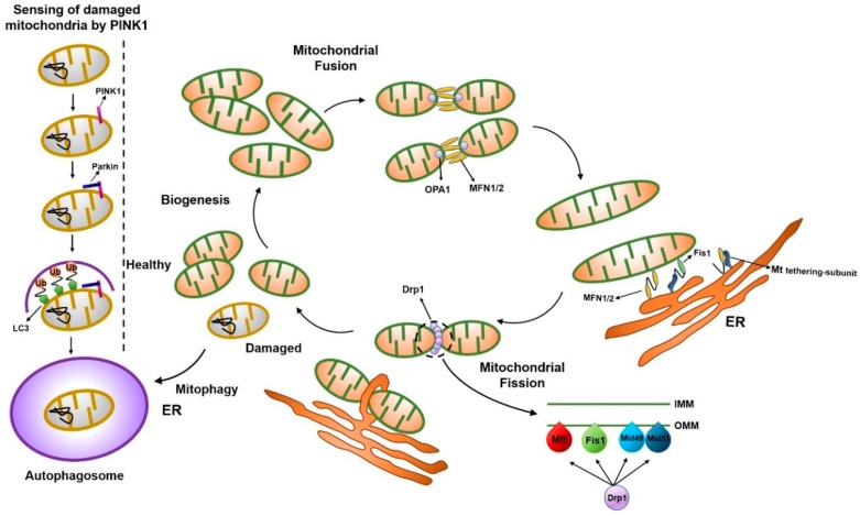
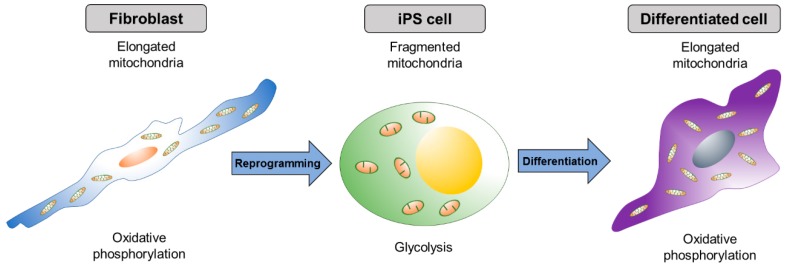
Mitochondria are highly dynamic organelles that continuously change their shape. Their main function is adenosine triphosphate (ATP) production; however, they are additionally involved in a variety of cellular phenomena, such as apoptosis, cell cycle, proliferation, differentiation, reprogramming, and aging. The change in mitochondrial morphology is closely related to the functionality of mitochondria. Normal mitochondrial dynamics are critical for cellular function, embryonic development, and tissue formation. Thus, defects in proteins involved in mitochondrial dynamics that control mitochondrial fusion and fission can affect cellular differentiation, proliferation, cellular reprogramming, and aging. Here, we review the processes and proteins involved in mitochondrial dynamics and their various associated cellular phenomena.
Keywords: differentiation; fission; fusion; mitochondria; mitochondrial dynamics; pluripotency.
Conflict of interest statement
The authors declare no conflict of interest.
Figures
References
-
- Zuchner S., Mersiyanova I.V., Muglia M., Bissar-Tadmouri N., Rochelle J., Dadali E.L., Zappia M., Nelis E., Patitucci A., Senderek J., et al. Mutations in the mitochondrial GTPase mitofusin 2 cause Charcot-Marie-Tooth neuropathy type 2A. Nat. Genet. 2004;36:449–451. doi: 10.1038/ng1341. - DOI - PubMed
Publication types
MeSH terms
Grants and funding
LinkOut - more resources
Full Text Sources
Other Literature Sources
Medical
