. 2018 Dec 18;19(1):12.
doi: 10.1186/s12858-018-0101-0.

Soluble expression of recombinant midgut zymogen (native propeptide) proteases from the Aedes aegypti Mosquito Utilizing E. coli as a host

Affiliations

Soluble expression of recombinant midgut zymogen (native propeptide) proteases from the Aedes aegypti Mosquito Utilizing E. coli as a host

James T Nguyen et al. BMC Biochem. .

Abstract

Background: Studying proteins and enzymes involved in important biological processes in the Aedes aegypti mosquito is limited by the quantity that can be directly isolated from the mosquito. Adding to this difficulty, digestive enzymes (midgut proteases) involved in metabolizing blood meal proteins require a more oxidizing environment to allow proper folding of disulfide bonds. Therefore, recombinant techniques to express foreign proteins in Escherichia coli prove to be effective in producing milligram quantities of the expressed product. However, with the most commonly used strains having a reducing cytoplasm, soluble expression of recombinant proteases is hampered. Fortunately, new E. coli strains with a more oxidizing cytoplasm are now available to ensure proper folding of disulfide bonds.

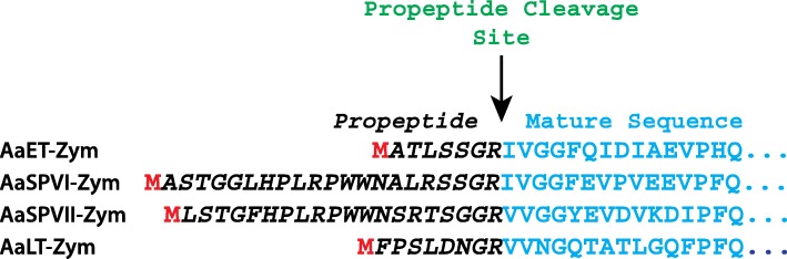
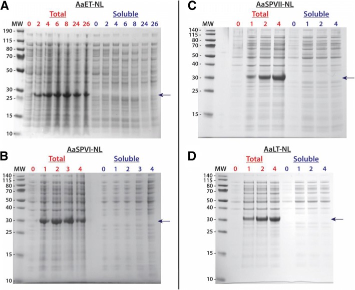
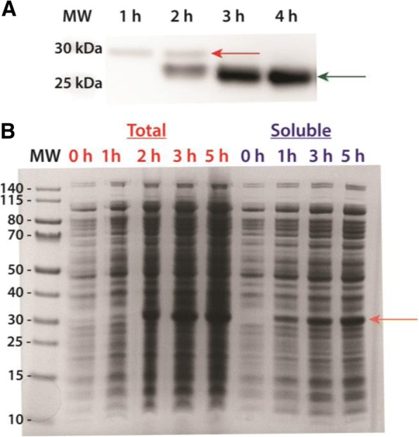
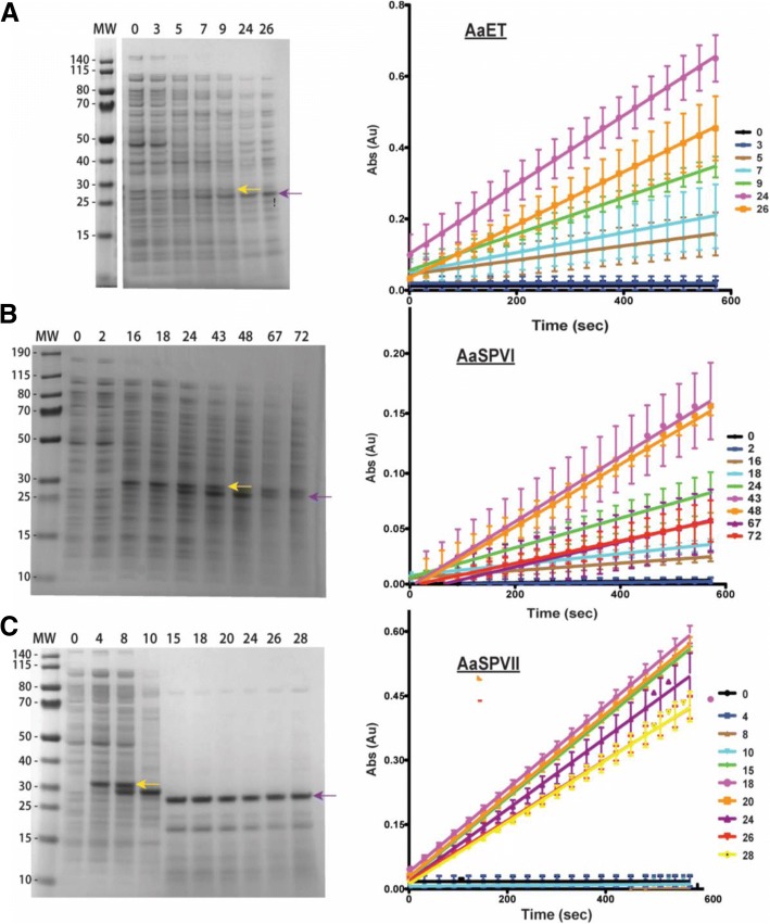
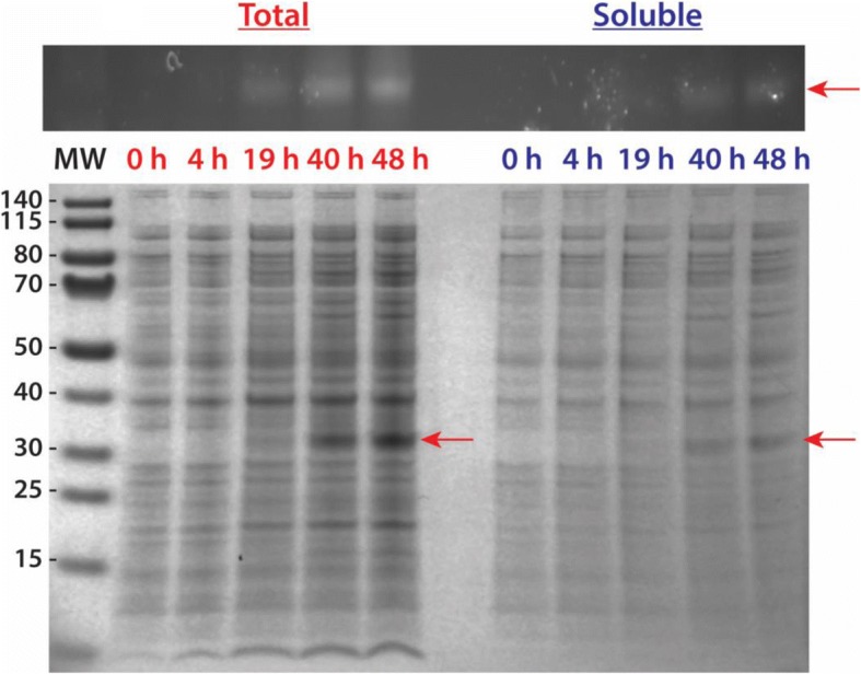
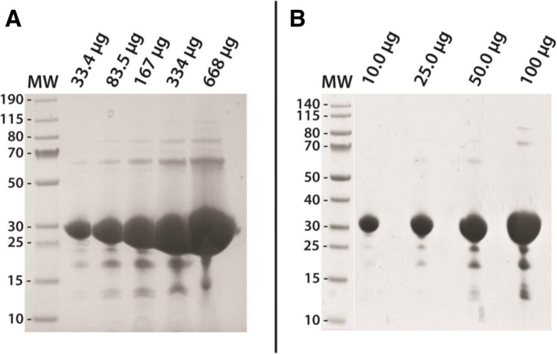
Results: Utilizing an E. coli strain with a more oxidizing cytoplasm (SHuffle® T7, New England Biolabs) and changes in bacterial growth temperature has resulted in the soluble expression of the four most abundantly expressed Ae. aegypti midgut proteases (AaET, AaSPVI, AaSPVII, and AaLT). A previous attempt of solubly expressing the full-length zymogen forms of these proteases with the leader (signal) sequence and a modified pseudo propeptide with a heterologous enterokinase cleavage site led to insoluble recombinant protein expression. In combination with the more oxidizing cytoplasm, and changes in growth temperature, helped improve the solubility of the zymogen (no leader) native propeptide proteases in E. coli. Furthermore, the approach led to autocatalytic activation of the proteases during bacterial expression and observable BApNA activity. Different time-points after bacterial growth induction were tested to determine the time at which the inactive (zymogen) species is observed to transition to the active form. This helped with the purification and isolation of only the inactive zymogen forms using Nickel affinity.

Conclusions: The difficulty in solubly expressing recombinant proteases in E. coli is caused by the native reducing cytoplasm. However, with bacterial strains with a more oxidizing cytoplasm, recombinant soluble expression can be achieved, but only in concert with changes in bacterial growth temperature. The method described herein should provide a facile starting point to recombinantly expressing Ae. aegypti mosquito proteases or proteins dependent on disulfide bonds utilizing E. coli as a host.

Keywords: Aedes aegypti; Disulfide bond/bridge; Escherichia coli; Midgut; Proteases; Recombinant protein; Soluble expression; Zymogen.


Conflict of interest statement

Ethics approval and consent to participate

Not applicable.

Consent for publication

Not applicable.

Competing interests

The authors declare that they have no competing interests.

Publisher’s Note

Springer Nature remains neutral with regard to jurisdictional claims in published maps and institutional affiliations.

Figures

Fig. 1
Fig. 1
Amino acid sequences of Ae. aegypti midgut zymogen (no leader) proteases. In order to improve solubility of recombinant proteases, the leader (signal) sequence was removed to produce the no leader zymogen as shown. Since the genes were cloned into the pET28a vector (utilizing the NdeI restriction site, CATATG), the resulting recombinant proteases will contain an N-terminal Methionine (shown in red), as well as the his6-tag linker (MGSSHHHHHHSSGLVPRGSH) upstream of the Met group (shown in red). The arrow points to the propeptide cleavage site required to activate the zymogen to the active mature protease
Fig. 2
Fig. 2
Initial attempt at solubly expressing recombinant midgut proteases in SHuffle® T7 Express Competent E. coli cells (NEB). For each growth experiment, TB media and a 30 °C growth temperature was used. Cells were induced with 0.1 mM IPTG when reaching the log phase (OD600nm ~ 0.5–1.0). Samples were collected at the given time points (in hours) and prepared for SDS-PAGE analysis. The MW ladder is in kilo-Daltons (kDa). In all cases, the arrow indicates where the expected soluble over-expressed protease should appear. However, all proteases under these conditions were expressed insolubly, only observed in the total samples. a 4–12% BIS-TRIS gel over-expression of AaET grown for a total of 26 h. The MW of the his6-tagged AaET-NL zymogen is ~ 27.0 kDa. b 12% BIS-TRIS gel over-expression of AaSPVI grown for a total of 4 h. The MW of the his6-tagged AaSPVI-NL zymogen is ~ 28.7 kDa. c 12% BIS-TRIS gel over-expression of AaSPVII grown for a total of 4 h. The MW of the his6-tagged AaSPVII-NL zymogen is ~ 28.7 kDa. d 12% BIS-TRIS gel over-expression of AaLT grown for a total of 4 h. The MW of the his6-tagged AaLT-NL zymogen is ~ 27.6 kDa
Fig. 3
Fig. 3
Soluble expression of recombinant AaET-NL and AaSPVII-NL zymogen proteases grown in TB media at 23 °C post-induction (induced with 0.1 mM IPTG). Plasmid constructs were transformed into SHuffle® T7 Express Competent E. coli cells (NEB). The MW ladder is in kilo-Daltons (kDa). a Western blot analysis utilizing an AaET-specific antibody of soluble samples collected from the growth and expression of AaET (a total of 4 h post-induction). The zymogen (inactive form of the protease) is observed in the first 2 h (MW ~ 27.0 kDa, red arrow), but a second species hypothesized to be the active mature form begins to appear at the two-hour time-point (MW ~ 22.4 kDa, green arrow) while the zymogen completely disappears by the third hour post-induction. b Large scale expression analysis of AaSPVII-zymogen grown for a total of 5 h post-induction. A single band at ~ 28.7 kDa (orange arrow) is observed to be increasing over time after induction with no observable band present in both the total and soluble pre-induction samples (t = 0 h)
Fig. 4
Fig. 4
SDS-PAGE analysis and BApNA activity assays of samples collected from small-scale growth experiments of SHuffle® E. coli cells (NEB) grown in TB media at 15 °C (induced with 0.1 mM IPTG). Samples were collected at the given time points (in hours). The MW ladder is in kilo-Daltons (kDa). a The gel represents the soluble expression of AaET-NL zymogen (MW ~ 27.0 kDa, yellow arrow), auto-activating to the active mature form (MW ~ 22.4 kDa, purple arrow). The presence of active AaET at 5 h post-induction correlates with an increase in BApNA activity, with maximal activity observed at the 24 h time-point (plot on the right). b The gel represents the soluble expression of AaSPVI-NL zymogen (MW ~ 28.7 kDa, yellow arrow), auto-activating to the active mature form (MW ~ 24.1 kDa, purple arrow). The presence of active AaSPVI at 16 h post-induction correlates with an increase in BApNA activity, with maximal activity observed at the 67 h time-point (plot on the right). c The gel represents the soluble expression of AaSPVII-NL zymogen (MW ~ 28.7 kDa, yellow arrow), auto-activating to the active mature form (MW ~ 24.2 kDa, purple arrow). The presence of active AaSPVII at 15 h post-induction correlates with an increase in BApNA activity, with maximal activity observed at the 18 h time-point (plot on the right). Unlike AaET and AaSPVI, AaSPVII expression results in a species that lies between the zymogen and active forms starting at 8 h post-induction and disappearing at 15 h. This species is an inactive form of AaSPVII since no detectable BApNA activity observed
Fig. 5
Fig. 5
Large-scale soluble expression of recombinant AaLT-NL zymogen protease grown in TB media at 10 °C (induced with 0.1 mM IPTG). Plasmid construct was transformed into SHuffle® T7 Express Competent E. coli cells (NEB). Samples were collected at the given time points (in hours). The MW ladder is in kilo-Daltons (kDa). Gel analysis of samples collected from the growth of AaLT was first visualized using InVision™ His-Tag In-Gel Stain (Invitrogen), which specifically chelates to and enhances the fluorescence of poly his-tagged proteins (top figure). The His-Tag stain is the positive identification that the bands expressed in the gel below are indeed the expression of soluble AaLT-zymogen (MW ~ 27.6 kDa, red arrows). The growth was extended beyond 24 h due to the 10 °C growth conditions, which helped in solubly expressing the protease, but also to increase bacterial cell density in order to obtain a large quantity of cell paste for purification
Fig. 6
Fig. 6
Final gel of Nickel purified recombinant zymogen (no leader) proteases grown and expressed at either 23 °C or 10 °C. In order to demonstrate that inactive zymogen proteases (with intact N-terminal his6-tag) can be isolated, AaSPVII grown at 23 °C (a) and AaLT grown at 10 °C (b) were purified. These samples are the post-dialysis concentrate of AaSPVI-NL and AaLT-NL dialyzed in 50 mM Sodium Acetate pH 5.2 + 2 mM DTT (buffer exchanged twice and set at 4 °C). Samples loaded on the gel are in micrograms (μg) and are increasing in order to show that the single step purification scheme led to a near homogenous sample with very little contaminants

Similar articles

Cited by

References

    1. Isoe J, Rascon AA, Jr, Kunz S, Miesfeld RL. Molecular genetic analysis of midgut serine proteases in Aedes aegypti mosquitoes. Insect Biochem Mol Biol. 2009;39(12):903–912. doi: 10.1016/j.ibmb.2009.10.008. - DOI - PMC - PubMed
    1. Monath TP. Dengue and yellow fever--challenges for the development and use of vaccines. N Engl J Med. 2007;357(22):2222–2225. doi: 10.1056/NEJMp0707161. - DOI - PubMed
    1. Schwartz O, Albert ML. Biology and pathogenesis of chikungunya virus. Nat Rev Microbiol. 2010;8(7):491–500. doi: 10.1038/nrmicro2368. - DOI - PubMed
    1. Casale TB, Teng MN, Morano JP, Unnasch T, Lockwood CJ. Zika virus: an emerging infectious disease with serious perinatal and neurologic complications. J Allergy Clin Immunol. 2018;141(2):482–490. doi: 10.1016/j.jaci.2017.11.029. - DOI - PubMed
    1. Elizondo-Quiroga D, Medina-Sanchez A, Sanchez-Gonzalez JM, Eckert KA, Villalobos-Sanchez E, Navarro-Zuniga AR, Sanchez-Tejeda G, Correa-Morales F, Gonzalez-Acosta C, Arias CF, et al. Zika virus in salivary glands of five different species of wild-caught mosquitoes from Mexico. Sci Rep. 2018;8(1):809. doi: 10.1038/s41598-017-18682-3. - DOI - PMC - PubMed

Publication types

MeSH terms

LinkOut - more resources