Review
. 2019 Feb 8;294(6):2098-2108.
doi: 10.1074/jbc.REV118.002804. Epub 2018 Dec 18.

The endoplasmic reticulum (ER) chaperone BiP is a master regulator of ER functions: Getting by with a little help from ERdj friends

Affiliations
Review

The endoplasmic reticulum (ER) chaperone BiP is a master regulator of ER functions: Getting by with a little help from ERdj friends

Kristine Faye R Pobre et al. J Biol Chem. .

Abstract

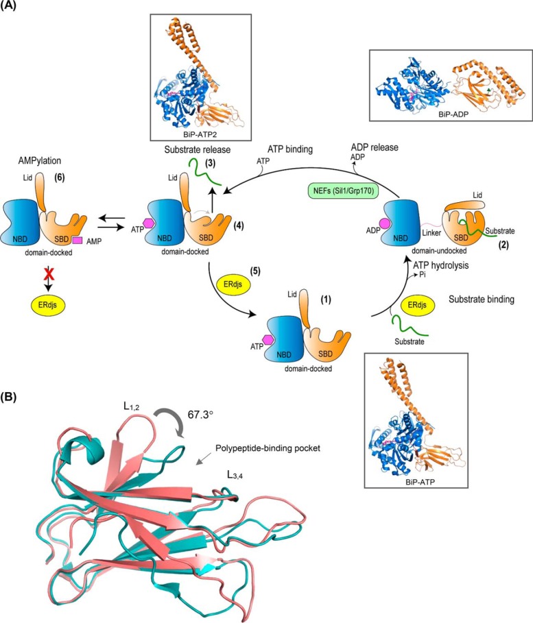
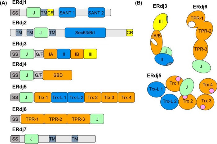
The endoplasmic reticulum (ER) represents the entry point into the secretory pathway where nascent proteins encounter a specialized environment for their folding and maturation. Inherent to these processes is a dedicated quality-control system that detects proteins that fail to mature properly and targets them for cytosolic degradation. An imbalance in protein folding and degradation can result in the accumulation of unfolded proteins in the ER, resulting in the activation of a signaling cascade that restores proper homeostasis in this organelle. The ER heat shock protein 70 (Hsp70) family member BiP is an ATP-dependent chaperone that plays a critical role in these processes. BiP interacts with specific ER-localized DnaJ family members (ERdjs), which stimulate BiP's ATP-dependent substrate interactions, with several ERdjs also binding directly to unfolded protein clients. Recent structural and biochemical studies have provided detailed insights into the allosteric regulation of client binding by BiP and have enhanced our understanding of how specific ERdjs enable BiP to perform its many functions in the ER. In this review, we discuss how BiP's functional cycle and interactions with ERdjs enable it to regulate protein homeostasis in the ER and ensure protein quality control.

Keywords: ATPase; BiP; ER-localized DnaJ proteins; GRP78; endoplasmic reticulum (ER); endoplasmic reticulum–associated protein degradation (ERAD); heat shock protein (HSP); molecular chaperone; protein folding; protein misfolding; stress response; unfolded protein response (UPR).


Conflict of interest statement

The authors declare that they have no conflicts of interest with the contents of this article

Figures

Figure 1.
Figure 1.
A, BiP ATPase cycle. Step 1, in the ATP-bound form, the nucleotide-binding domain (NBD) (blue) and the substrate-binding domain (SBD) (orange), with its lid open, are docked to each other resulting to a form with high-substrate binding and release kinetics and low-substrate affinity. Step 2, upon ATP hydrolysis, the NBD and SBD become undocked, and the lid of the SBD closes providing a form that has high-substrate affinity but slow binding and release rates. This cycle is regulated by ER-localized DnaJ cofactors (ERdjs) that interact with unfolded proteins and transfer them to the ATP-bound form of BiP, while simultaneously triggering ATP hydrolysis. Step 3, substrate is released with the help of nucleotide-exchange factors (NEFs) that stimulate the release of ADP. Step 4, binding of ATP causes a conformational change in the SBD resulting in a more tightly compacted conformation that is thought to “squeeze” the substrate out. Step 5, interaction with ERdjs reorders the polypeptide-binding pocket of the BiP–ATP2 SBD, readying it to interact with another substrate. Step 6, BiP is post-translationally modified through AMPylation, and this causes the protein to be inactive. AMPylated BiP adopts a “domain-docked” structure similar to that of the ATP-bound state even in the apo- or ADP-bound state and is unable to interact productively with ERdjs. Ribbon representations of the structures (insets): ATP-bound BiP with the polypeptide-binding pocket open (BiP-ATP) (PDB 5e84 (8)); ADP-bound BiP from the structures of the isolated NBD (PDB 5evz (102)); SBD (PDB 5e85 (8)); and ATP-bound BiP with the polypeptide-binding pocket fully closed (BiP-ATP2) (PDB 6asy (8)). B, overlay of the SBD of BiP–ATP and BiP–ATP2. Comparison of SBDβ of BiP–ATP (deep salmon, PDB 5e84) and BiP–ATP2 (teal, PDB 6asy) with their superposition based on Cαs of β3–β7. The peptide-binding loops L1, 2 and L3, 4 are shown. The main difference of the two conformations is that the L1, 2 in BiP–ATP2 is rotated more than 60° compared with that of BiP–ATP resulting in a fully closed polypeptide-binding pocket.
Figure 2.
Figure 2.
Domain structure of ERdj family members. A, domain arrangements of primary sequences of each of the known ERdj proteins. Abbreviations used are as follows: SS, signal sequence; J, J domain; TM, transmembrane region; CR, charged amino acid region; SANT, SANT domain; Sec63/Brl, Sec63/Brl domain; G/F, glycine/phenylalanine-rich flexible linker region; IA and IB, bifurcated substrate-binding domain; II, cysteine-rich domain; III, dimerization domain; SBD, substrate-binding domain; Trx, thioredoxin domains; Trx-L, thioredoxin-like, enzymatically inactive domain; TPR, tetratricopeptide repeat domain (each containing three subdomains). B, cartoons representing orientation of the various domains of ERdj5 (72, 73) and ERdj6 (93, 94) based upon known structures. ERdj3 layout was derived from a combination of the solved structure of domain I–III of the yeast homologue Ydj1 (38, 39) and negative strain EM of ERdj3 showing the relative orientation of the J domain (43). The structures of ERdj1, -2, -4, and -7 are unsolved. The following functional regions are also indicated: ERdj3 and ERdj6 substrate-binding sites (white), ERdj3 dimerization/tetramerization regions within domains III and II respectively (blue-gray), and ERdj5's redox-active Trx motifs (pink).

References

    1. Ellgaard L., and Helenius A. (2003) Quality control in the endoplasmic reticulum. Nat. Rev. Mol. Cell Biol. 4, 181–191 10.1038/nrm1052 - DOI - PubMed
    1. Braakman I., and Bulleid N. J. (2011) Protein folding and modification in the mammalian endoplasmic reticulum. Annu. Rev. Biochem. 80, 71–99 10.1146/annurev-biochem-062209-093836 - DOI - PubMed
    1. Mayer M. P., and Kityk R. (2015) Insights into the molecular mechanism of allostery in Hsp70s. Front. Mol. Biosci. 2, 58 - PMC - PubMed
    1. Mayer M. P. (2018) Intra-molecular pathways of allosteric control in Hsp70s. Philos. Trans. R. Soc. Lond. B Biol. Sci. 373, 20170183 10.1098/rstb.2017.0183 - DOI - PMC - PubMed
    1. Misselwitz B., Staeck O., and Rapoport T. A. (1998) J proteins catalytically activate Hsp70 molecules to trap a wide range of peptide sequences. Mol. Cell 2, 593–603 10.1016/S1097-2765(00)80158-6 - DOI - PubMed

Publication types

MeSH terms