. 2018 Dec 18;11(561):eaal2171.
doi: 10.1126/scisignal.aal2171.

5-oxoETE triggers nociception in constipation-predominant irritable bowel syndrome through MAS-related G protein-coupled receptor D

Affiliations

5-oxoETE triggers nociception in constipation-predominant irritable bowel syndrome through MAS-related G protein-coupled receptor D

Tereza Bautzova et al. Sci Signal. .

Abstract

Irritable bowel syndrome (IBS) is a common gastrointestinal disorder that is characterized by chronic abdominal pain concurrent with altered bowel habit. Polyunsaturated fatty acid (PUFA) metabolites are increased in abundance in IBS and are implicated in the alteration of sensation to mechanical stimuli, which is defined as visceral hypersensitivity. We sought to quantify PUFA metabolites in patients with IBS and evaluate their role in pain. Quantification of PUFA metabolites by mass spectrometry in colonic biopsies showed an increased abundance of 5-oxoeicosatetraenoic acid (5-oxoETE) only in biopsies taken from patients with IBS with predominant constipation (IBS-C). Local administration of 5-oxoETE to mice induced somatic and visceral hypersensitivity to mechanical stimuli without causing tissue inflammation. We found that 5-oxoETE directly acted on both human and mouse sensory neurons as shown by lumbar splanchnic nerve recordings and Ca2+ imaging of dorsal root ganglion (DRG) neurons. We showed that 5-oxoETE selectively stimulated nonpeptidergic, isolectin B4 (IB4)-positive DRG neurons through a phospholipase C (PLC)- and pertussis toxin-dependent mechanism, suggesting that the effect was mediated by a G protein-coupled receptor (GPCR). The MAS-related GPCR D (Mrgprd) was found in mouse colonic DRG afferents and was identified as being implicated in the noxious effects of 5-oxoETE. Together, these data suggest that 5-oxoETE, a potential biomarker of IBS-C, induces somatic and visceral hyperalgesia without inflammation in an Mrgprd-dependent manner. Thus, 5-oxoETE may play a pivotal role in the abdominal pain associated with IBS-C.


Conflict of interest statement

Competing interests: The authors declare that they have no competing interests.

Figures

Fig. 1
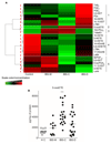
Fig. 1. Quantification of PUFA metabolites in mucosa of IBS patients.
(A) Heat-map of PUFA metabolites quantified by liquid chromatography-tandem mass spectrometry. Data are shown in a matrix format: each row represents a single PUFA metabolite, and each column represents a subgroup of patients. Each color patch represents the normalized quantity of PUFA metabolites (row) in a subgroup of patients (column), with a continuum of quantity from bright green (lowest) to bright red (highest). The pattern and length of the branches in the dendrograms reflect the relatedness of the PUFA metabolites. The dashed red line is the dendrogram distance used to cluster PUFA metabolites. (B) 5-oxoETE quantified by liquid chromatography-tandem mass spectrometry in the mucosa of HCs (white circle) and patients with the indicated type of IBS (black circle). Data are expressed in pg/mg protein and presented as means ± SEM of 10 to 20 biopsies per group. Statistical analysis was performed using Kruskal-Wallis analysis of variance and subsequent Dunn’s post hoc test. ***P < 0.001 compared to the HC group.
Fig. 2
Fig. 2. 5-oxoETE induces somatic and visceral hypersensitivity in vivo.
(A to E) Eight-week-old male C57BL/6J mice were subcutaneously injected with either HBSS (white circle) or 5-oxoETE (black circle) into hind footpads. (A) Somatic pain was monitored using the von Frey test at the indicated times after injection with 10 μM 5-oxoETE or HBSS. Data are means of three independent experiments with 5 mice per group. Errors bars indicate SEM. (B) The Von Frey test was performed 30 min after injection of the indicated concentrations of 5-oxoETE. Data are means ± SEM of two experiments with six mice per group. (C) Mouse paw tissue samples were stained with H&E 6 hours after the administration of HBSS or 100 μM 5-oxoETE as indicated. Images are representative of two experiments with 5 mice per group. (D) Visceromotor response (VMR) to increasing pressures of colorectal distension before and 30 min after intracolonic administration of 10μM 5-oxoETE (black bars) or vehicle (40% ethanol; white bars). Data are means ± SEM of two experiments with 10 mice per group and are relative to the baseline recorded before treatment. (E) Colon tissue samples stained with H&E from mice treated with 40% ethanol or 10 μM 5-oxoETE as indicated. Images are representative of two experiments with 5 mice per group. Statistical analysis was performed using Kruskal-Wallis analysis of variance and subsequent Dunn’s post hoc test. **P < 0.01, ***P < 0.001, compared to control mice.
Fig. 3
Fig. 3. 5-oxoETE induces lumbar splanchnic nerve firing.
(A) Example of a teased-fiber recording showing the lumbar splanchnic (that is, colon-innervating) nerve response to ring application (7 min) of 5-oxoETE in mouse serosal afferents. Arrows indicate application and removal of 5-oxoETE. Data are representative of eight experiments in which 5-oxoETE elicited nerve discharge above baseline from a total of 21 teased fibers isolated from 7 mice. (B) Mean change in firing/s in serosal receptive fields in response to 5-oxo-ETE compared with the response to vehicle (Krebs buffer). Data are means ± SEM of 8 teased-fiber recordings (N = 7 mice) for 5-oxoETE and 5 teased-fiber recordings (N = 5 mice) for vehicle. Statistical analysis was performed using a Mann-Whitney t-test. **P < 0.01 compared to vehicle. (C) Proportion of responses in lumbar splanchnic afferents to application of 5-oxoETE (n, number of teased-fiber recordings; N, number of mice).
Fig. 4
Fig. 4. 5-oxoETE induces an increase in [Ca2+]i in sensory neurons through a GPCR.
(A) Representative trace of Ca2+ flux experiments in sensory neurons incubated in the absence of extracellular Ca2+/Mg2+ and exposed to 50 μM 5-oxoETE or vehicle (HBSS). (B) Ca2+ flux measurements in mouse sensory neurons exposed to the indicated concentrations of 5-oxoETE (black circle) or to vehicle (HBSS; white circle). Data are means + SEM of seven independent experiments with 3 wells per condition and 60 to 80 neurons per well. (C) Amplitude of [Ca2+]i (ΔF/F; left) in human sensory neurons and the percentage of responding neurons (right) exposed to the indicated concentrations of 5-oxoETE (black bar) or to vehicle (HBSS; white bar). Data are means ± SEM of three independent experiments with 3 wells per condition and 20 to 53 neurons per well. (D) Percentages of isolectin B4–positive (IB4+) and IB4-negative (IB4-) mouse sensory neurons that responded to 10 μM 5-oxoETE (black bars) or HBSS (white bars). Data are means ± SEM of three independent experiments with 3 wells per condition and 60 to 80 neurons per well. (E) Effects of 30-min incubation with 10 μM U73122 (PLC inhibitor) or overnight incubation with PTX (250 ng/ml) on 5-oxoETE–induced Ca2+ mobilization in mouse sensory neurons. Data are means ± SEM of five independent experiments with 3 wells per condition and 60 to 80 neurons per well. Statistical analysis was performed using Kruskal-Wallis analysis of variance and subsequent Dunn’s post hoc test. *P < 0.05, **P < 0.01, ***P < 0.001 compared to HBSS.
Fig. 5
Fig. 5. Expression of Mrgprd in sensory neurons.
(A) Expression of Mrgprd (red) and Trpv1 (blue) mRNA transcripts as detected by single-cell qRT-PCR analysis (middle) of retrogradely labelled mouse colonic sensory neurons (left). Pie chart representation (right) of the expression (dark color) or not (light color) of Mrgprd and Trpv1 mRNA in Fast Blue (FB)-positive neurons. Each segment represents a single colonic sensory neuron. (B) Representative images of GFP (green), CGRP-immunoreactivity (red), and FB labelling (blue) in a T13 DRG from a MrgprdEGFP mouse in which FB was injected into the colon. Scale bar, 50 μm. Inset images are magnifications of the boxed areas in the largest images. (C and D) Expression of Mrgprd by immunostaining in a whole human T11 DRG (C) and in a primary culture of human sensory neurons using confocal microscopy (D). Pie chart representation of the immunoreactivity (dark color) or not (light color) of Mrgprd in Pgp9.5-positive neurons. Each segment represents a single sensory neuron. Scale bars, 10 μm. Images are representative of 2 experiments with 10 slides per experiment (C) and of 5 experiments with 2 wells per experiment (D).
Fig. 6
Fig. 6. Mrgprd expression is required for the intracellular Ca2+ mobilization and hypersensitivity induced by 5-oxoETE.
(A) Left: Representative image of sensory neurons transfected with shRNA (red) and containing the Ca2+ indicator Fluo4 (green). Right: Percentage of sensory neurons expressing control shRNA or Mrgprd-specific shRNA that responded to HBSS or 10 μM 5-oxoETE. Data are means ± SEM of six independent experiments with 3 wells per conditions and 10 to 32 analyzed neurons per well. (B) Percentage of responding neurons (left) and amplitude of intracellular Ca2+ mobilization (ΔF/F; right) in mouse sensory neurons from Mrgprd-deficient mice exposed to vehicle (HBSS; white bar), 10 μM 5-oxoETE (black bar), or to a mixture of GPCR agonists (GPCR Mix: bradykinin, serotonin, and histamine, 10 μM each; gray bar). Data are means ± SEM of four independent experiments with 3 wells per condition and 20 to 50 neurons per well. (C) Effects of the indicated concentrations of 5-oxoETE and of 1 mM β-alanine (positive control) on the amplitude (ΔF/F) of Ca2+ mobilization in HEK cells transiently transfected with plasmid expressing Mrgprd or with an empty vector as a control. Data are means ± SEM of eight independent experiments with 3 wells per condition. (D) Visceromotor response (VMR) in Mrgprd-deficient mice in response to increasing pressures of colorectal distension before (baseline; white circle) and 30 min after intracolonic administration of 10 μM 5-oxoETE (black circle). Data are means + SEM of two experiments of 7 mice per experiment. Statistical analysis was performed using Kruskal-Wallis analysis of variance and subsequent Dunn’s post hoc test. In (A), **P < 0.01 compared to the control shRNA/HBSS group; In (B), **P < 0.01 compared to the HBSS group; In (C), *P < 0.05, **P < 0.01, ***P < 0.001 compared to the corresponding CHO empty vector group.

References

    1. Mearin F, Lacy BE, Chang L, Chey WD, Lembo AJ, Simren M, Spiller R. Bowel Disorders. Gastroenterology. 2016 - PubMed
    1. Enck P, Aziz Q, Barbara G, Farmer AD, Fukudo S, Mayer EA, Niesler B, Quigley EM, Rajilic-Stojanovic M, Schemann M, Schwille-Kiuntke J, et al. Irritable bowel syndrome. Nat Rev Dis Primers. 2016;2 16014. - PMC - PubMed
    1. Ohman L, Simren M. Pathogenesis of IBS: role of inflammation, immunity and neuroimmune interactions. Nat Rev Gastroenterol Hepatol. 2010;7:163–173. - PubMed
    1. Cenac N, Bautzova T, Le Faouder P, Veldhuis NA, Poole DP, Rolland C, Bertrand J, Liedtke W, Dubourdeau M, Bertrand-Michel J, Zecchi L, et al. Quantification and Potential Functions of Endogenous Agonists of Transient Receptor Potential Channels in Patients With Irritable Bowel Syndrome. Gastroenterology. 2015;149:433–444 e437. - PubMed
    1. Barbara G, Wang B, Stanghellini V, De G R, Cremon C, Di N G, Trevisani M, Campi B, Geppetti P, Tonini M, Bunnett NW, et al. Mast cell-dependent excitation of visceral-nociceptive sensory neurons in irritable bowel syndrome. Gastroenterology. 2007;132:26–37. - PubMed

Publication types

MeSH terms

LinkOut - more resources