. 2018 Dec 18;9(6):e02353-18.
doi: 10.1128/mBio.02353-18.

Transcriptional Profiling of Patient Isolates Identifies a Novel TOR/Starvation Regulatory Pathway in Cryptococcal Virulence

Affiliations

Transcriptional Profiling of Patient Isolates Identifies a Novel TOR/Starvation Regulatory Pathway in Cryptococcal Virulence

Yoon-Dong Park et al. mBio. .

Abstract

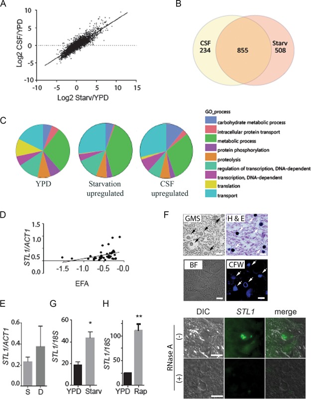
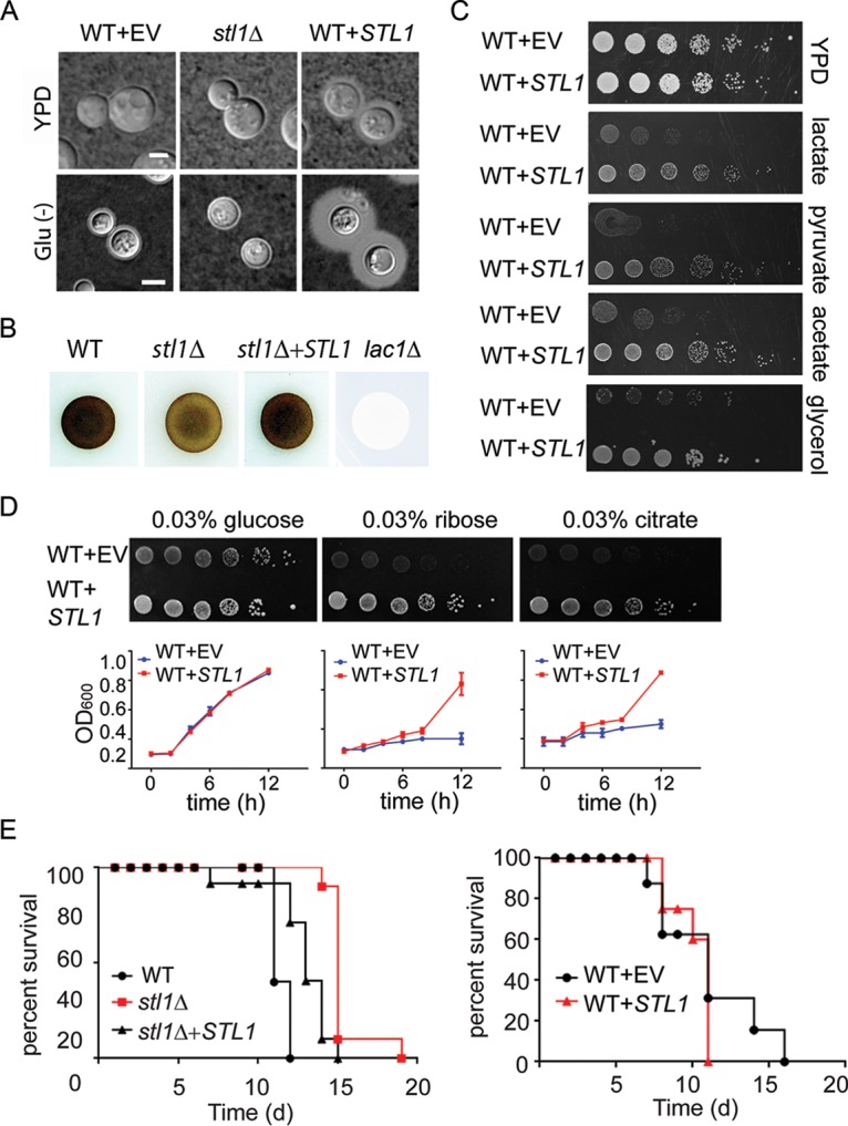
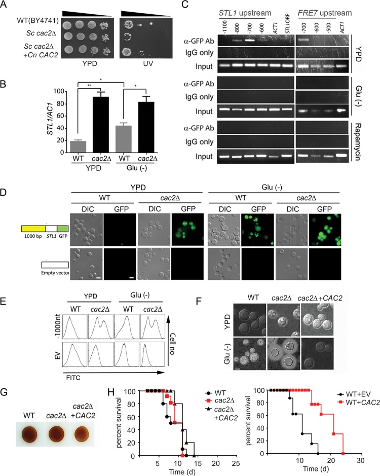
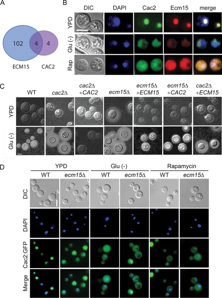
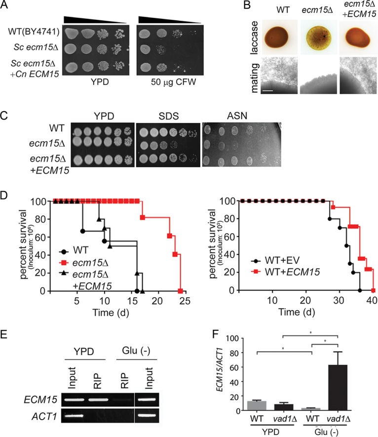
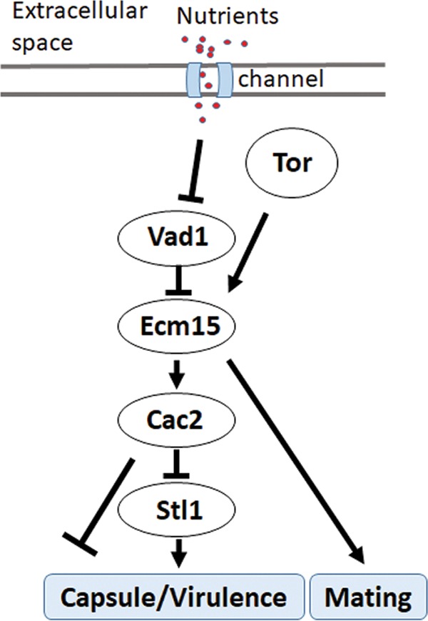
Human infection with Cryptococcus causes up to a quarter of a million AIDS-related deaths annually and is the most common cause of nonviral meningitis in the United States. As an opportunistic fungal pathogen, Cryptococcus neoformans is distinguished by its ability to adapt to diverse host environments, including plants, amoebae, and mammals. In the present study, comparative transcriptomics of the fungus within human cerebrospinal fluid identified expression profiles representative of low-nutrient adaptive responses. Transcriptomics of fungal isolates from a cohort of HIV/AIDS patients identified high expression levels of an alternative carbon nutrient transporter gene, STL1, to be associated with poor early fungicidal activity, an important clinical prognostic marker. Mouse modeling and pathway analysis demonstrated a role for STL1 in mammalian pathogenesis and revealed that STL1 expression is regulated by a novel multigene regulatory mechanism involving the CAC2 subunit of the chromatin assembly complex 1, CAF-1. In this pathway, the global regulator of virulence gene VAD1 was found to transcriptionally regulate a cryptococcal homolog of a cytosolic protein, Ecm15, in turn required for nuclear transport of the Cac2 protein. Derepression of STL1 by the CAC2-containing CAF-1 complex was mediated by Cac2 and modulated binding and suppression of the STL1 enhancer element. Derepression of STL1 resulted in enhanced survival and growth of the fungus in the presence of low-nutrient, alternative carbon sources, facilitating virulence in mice. This study underscores the utility of ex vivo expression profiling of fungal clinical isolates and provides fundamental genetic understanding of saprophyte adaption to the human host.IMPORTANCECryptococcus is a fungal pathogen that kills an estimated quarter of a million individuals yearly and is the most common cause of nonviral meningitis in the United States. The fungus is carried in about 10% of the adult population and, after reactivation, causes disease in a wide variety of immunosuppressed individuals, including the HIV infected and patients receiving transplant conditioning, cancer therapy, or corticosteroid therapy for autoimmune diseases. The fungus is widely carried in the soil but can also cause infections in plants and mammals. However, the mechanisms for this widespread ability to infect a variety of hosts are poorly understood. The present study identified adaptation to low nutrients as a key property that allows the fungus to inhabit these diverse environments. Further studies identified a nutrient transporter gene, STL1, to be upregulated under low nutrients and to be associated with early fungicidal activity, a marker of poor clinical outcome in a cohort of HIV/AIDS patients. Understanding molecular mechanisms involved in adaptation to the human host may help to design better methods of control and treatment of widely dispersed fungal pathogens such as Cryptococcus.

Keywords: CAC2; CAF-1; Cryptococcus neoformans; STL1; TOR pathway; VAD1.


Figures

FIG 1
FIG 1
Expression profiling demonstrates a concordance of cryptococcal genes upregulated in fungus-infected human cerebrospinal fluid and starvation. (A) C. neoformans strain H99 was grown to the mid-log phase in YPD and then subjected to starvation (Starv) in asparagine salts without glucose or sterile cerebrospinal fluid (CSF) at 37°C for 3 h. RNA was recovered and subjected to microarray analysis, and values were expressed as log2 ratios of expression in CSF/YPD. n = 2 for each condition. (B) Venn diagram of genes upregulated >2-fold and adj P < 0.05 after incubation in fungus-infected CSF or under starvation conditions. (C) GO biological process term occurrence among genes upregulated under the indicated conditions. (D) Transcriptional ratio of STL1/ACT1 determined by qRT-PCR of patient isolates compared to EFA (n = 46; regression analysis, n − 2 degrees of freedom [df], P < 0.05). (E) Expression ratios of STL1/ACT1 in survivors at 10 weeks (S [n = 35]) versus those who died (D [n = 11]). (F) Sections of a brain autopsy specimen were obtained from a 42-year-old man with HIV/AIDS who died of severe and diffuse C. neoformans infection and were visualized by bright-field microscopy (BF) or differential interference contrast microscopy (DIC) or stained with Gomori methenamine silver stain (GMS), hematoxylin and eosin (H&E stain), calcofluor white (CFW), or C3-fluorescein-labeled oligonucleotide probes for STL1 transcripts (STL1) by fluorescent in situ hybridization in the presence (+) or absence (−) of RNase A treatment. Arrows point to stained yeast cells. Bar = 10 µm. (G and H) Expression levels of STL1/18S rRNA under mid-log growth in YPD or 3 h of incubation under starvation conditions or in the presence of 5 µM rapamycin (Rap). n = 3 independent experiments. *, P < 0.05, and **, P < 0.01, by Student’s t test. Error bars represent standard deviation.
FIG 2
FIG 2
STL1 is required for growth on alternative carbon substrates, capsule formation, laccase activity, and mammalian virulence. (A) Indicated strains were incubated on YPD or ASN without glucose (Glu−) agar for 3 days at 30°C and examined by India ink microscopy. Bar = 5 µm. (B) The indicated cells were inoculated on ASN containing 100 mg/ml norepinephrine to assay production of melanin pigment by laccase. (C) The indicated strains were diluted to an A600 of 1.0, and 1:5 serial dilutions (5 μl) were plated on YPD or ASN containing the indicated substrates (0.03%) and incubated at 30°C for 5 days. (D) In the upper panels, the indicated strains were diluted to an A600 of 1.0, and 1:5 serial dilutions (5 μl) were plated on ASN containing the indicated substrates (0.03%) and incubated at 30°C for 7 days. In the lower panels, the indicated strains were cultured with ASN broth containing the indicated substrates (0.03%), and optical density at 600 nm (OD600) was calculated at the indicated time points. (E) Mice were inoculated by tail vein (106 of the indicated cells), and progress was followed until the mice were moribund. n = 10 mice per group. P < 0.0001 (left panel) and P = 0.5 (right panel) by log rank test.
FIG 3
FIG 3
Cac2 is a repressor of STL1 and capsule and overexpression reduces virulence in mice. (A) The indicated strains were diluted to an A600 of 1.0, and 1:5 serial dilutions (5 μl) were plated on YPD or on YPD with UV irradiation (UV) for 1 min and incubated at 30°C for 3 days. (B) qRT-PCR of STL1 of indicated strains grown to the mid-log phase in YPD and ASN without glucose (Glu−) media. n = 3 independent experiments. *, P < 0.05, and **, P < 0.01, by Student’s t test. (C) Chromatin immunoprecipitation (ChIP) of a Cac2-promoter complex. Nuclear extract from induced cryptococcal cells expressing a Cac2-GFP fusion protein was immunoprecipitated using an anti-GFP antibody (α-GFP Ab) or control IgG (IgG only) and assayed by PCR for the presence of the indicated regions of STL1, FRE7 promoter, or control promoter sequences of ACT1. (D and E) Effects of serial deletion of the STL1 5′-promoter (−1000 from the transcript start site) on basal transcriptional activity in C. neoformans. Linear constructs containing STL1 ORF with indicated regions of the STL1 5′-promoter–GFP fusion gene were transformed into WT or cac2Δ strains of C. neoformans, integration was confirmed by Southern blotting, and cells were grown to the mid-log phase in YPD or ASN salts without glucose (Glu−), and the population was subjected flow cytometry or microscopy (DIC or GFP fluorescence microscopy). Cells transformed with empty vector were used as control. Bar = 5 μm. (F) The indicated strains were inoculated onto the indicated media, recovered, and visualized by India ink microscopy. (G) Indicated cells were inoculated on ASN containing 100 mg/ml norepinephrine to assay production of melanin pigment by laccase. (H) Mice were inoculated by tail vein (106 cells of the indicated strains), and progress was followed until mice were moribund. n = 10 mice per group. P = 0.7 (left panel) and P < 0.0001 (right panel) by log rank test.
FIG 4
FIG 4
Cac2 and Ecm15 shares overlapping expression profiles and undergoes nutrient- and TOR-dependent nuclear localization. (A) Venn diagram of genes showing >2× increased transcription relative to WT and adj P < 0.05 after deletion of ECM15 or CAC2. (B) C. neoformans cells expressing Cac2-GFP or Ecm15-mCherry were incubated in YPD, ASN without glucose (Glu−), or YPD containing rapamycin and observed by fluorescence (DAPI, Cac2, or Ecm15) or differential interference contrast (DIC) microscopy. Bar = 5 µm. (C) Indicated strains were incubated on YPD or ASN without glucose (Glu−), recovered, and observed by India ink microsocopy. (D) Cells expressing Cac2-GFP in wild-type (WT) or ecm15Δ strains were incubated in YPD ASN without glucose (Glu−) or YPD containing rapamycin and observed by fluorescence or by DIC microscopy. Bar = 5 µm.
FIG 5
FIG 5
VAD1 is a repressor of ECM15, a regulator of capsule, laccase activity, and virulence. (A) The indicated strains were diluted to an A600 of 1.0, and 1:5 serial dilutions (5 μl) were plated on YPD medium or YPD containing 50 μg/ml of calcofluor white (CFW) and incubated at 30°C for 3 days. (B) The indicated strains were inoculated on ASN containing 100 mg/ml norepinephrine and observed after 2 days at 37°C (upper panel). Indicated MATα strains were coincubated with a MATa mating partner (strain KN99) on nitrogen-limiting mating medium (V8 agar) for 3 weeks at room temperature. The edges of the mating mixtures were photographed (40×). Bar = 500 µm (lower panel). (C) The indicated strains were diluted to an A600 of 1.0, and 1:5 serial dilutions (5 μl) were plated on YPD, ASN, or YPD containing SDS and incubated at 30°C for 3 to 7 days. (D) Mice were inoculated by tail vein (106 [left] or 103 [right] of the indicated strains), and progress was followed until they were moribund. n = 10 mice per group. P< 0.0001 (left panel) and P < 0.01 (right panel) by log rank test. (E) Lysates from cells expressing a c-myc-tagged Vad1 fusion protein incubated under the indicated conditions were immunoprecipitated followed by RT-PCR/gel electrophoresis using primers for the indicated gene transcripts. (F) Steady-state transcript levels of ECM15 from indicated strains grown to the mid-log phase in YPD or ASN salts without glucose (Glu−). n = 3 independent experiments. *; P < 0.05 by Student’s t test.
FIG 6
FIG 6
Scheme of starvation regulation of STL1 related to capsule formation and mammalian virulence.

References

    1. Park BJ, Wannemuehler KA, Marston BJ, Govender N, Pappas PG, Chiller TM. 2009. Estimation of the current global burden of cryptococcal meningitis among persons living with HIV/AIDS. AIDS 23:525–530. doi:10.1097/QAD.0b013e328322ffac. - DOI - PubMed
    1. Rajasingham R, Smith R, Park B, Jarvis J, Govender NP, Chiller T, Denning D, Loyse A, Boulware DR. 2017. Global burden of disease of HIV-associated cryptococcal meningitis: an updated analysis. Lancet Infect Dis 17:873–881. doi:10.1016/S1473-3099(17)30243-8. - DOI - PMC - PubMed
    1. Jarvis JN, Bicanic T, Loyse A, Namarika D, Jackson A, Nussbaum JC, Longley N, Muzoora C, Phulusa J, Taseera K, Kanyembe C, Wilson D, Hosseinipour MC, Brouwer AE, Limmathurotsakul D, White N, van der Horst C, Wood R, Meintjes G, Bradley J, Jaffar S, Harrison T. 2014. Determinants of mortality in a combined cohort of 501 patients with HIV-associated cryptococcal meningitis: implications for improving outcomes. Clin Infect Dis 58:736–745. doi:10.1093/cid/cit794. - DOI - PMC - PubMed
    1. Beardsley J, Wolbers M, Kibengo FM, Ggayi AB, Kamali A, Cuc NT, Binh TQ, Chau NV, Farrar J, Merson L, Phuong L, Thwaites G, Van Kinh N, Thuy PT, Chierakul W, Siriboon S, Thiansukhon E, Onsanit S, Supphamongkholchaikul W, Chan AK, Heyderman R, Mwinjiwa E, van Oosterhout JJ, Imran D, Basri H, Mayxay M, Dance D, Phimmasone P, Rattanavong S, Lalloo DG, Day JN, CryptoDex Investigators. 2016. Adjunctive dexamethasone in HIV-associated cryptococcal meningitis. N Engl J Med 374:542–554. doi:10.1056/NEJMoa1509024. - DOI - PMC - PubMed
    1. Williamson PR. 2013. Advancing translational immunology in HIV-associated cryptococcal meningitis. J Infect Dis 207:1793–1795. doi:10.1093/infdis/jit102. - DOI - PMC - PubMed

Publication types

MeSH terms

Substances