. 2018 Dec 3:(801):427-458.
doi: 10.3897/zookeys.801.22395. eCollection 2018.

Comparative ultrastructure of cells and cuticle in the anterior chamber and papillate region of Porcellioscaber (Crustacea, Isopoda) hindgut

Affiliations

Comparative ultrastructure of cells and cuticle in the anterior chamber and papillate region of Porcellioscaber (Crustacea, Isopoda) hindgut

Urban Bogataj et al. Zookeys. .

Abstract

Isopod hindgut consists of two anatomical and functional parts, the anterior chamber, and the papillate region. This study provides a detailed ultrastructural comparison of epithelial cells in the anterior chamber and the papillate region with focus on cuticle ultrastructure, apical and basal plasma membrane labyrinths, and cell junctions. Na+/K+-ATPase activity in the hindgut epithelial cells was demonstrated by cytochemical localisation. The main difference in cuticle ultrastructure is in the thickness of epicuticle which is almost as thick as the procuticle in the papillate region and only about one sixth of the thickness of procuticle in the anterior chamber. The apical plasma membrane in both hindgut regions forms an apical plasma membrane labyrinth of cytoplasmic strands and extracellular spaces. In the papillate region the membranous infoldings are deeper and the extracellular spaces are wider. The basal plasma membrane is extensively infolded and associated with numerous mitochondria in the papillate region, while it forms relatively scarce basal infoldings in the anterior chamber. The junctional complex in both hindgut regions consists of adherens and septate junctions. Septate junctions are more extensive in the papillate region. Na+/K+-ATPase was located mostly in the apical plasma membranes in both hindgut regions. The ultrastructural features of hindgut cuticle are discussed in comparison to exoskeletal cuticle and to cuticles of other arthropod transporting epithelia from the perspective of their mechanical properties and permeability. The morphology of apical and basal plasma membranes and localisation of Na+/K+-ATPase are compared with other arthropod-transporting epithelia according to different functions of the anterior chamber and the papillate region.

Keywords: cell junctions; digestive system; extracellular matrix; ion transporting epithelium; plasma membrane labyrinth.


Figures

Figure 1.
Figure 1.
Measurements in ImageJ/Fiji. A Measurements of cell width (line a), height (line b) and nucleus diameter (line c) B Measurements of cuticle and basal lamina thickness. Cuticle and basal lamina thickness (yellow lines) were measured at intersections of grid lines (blue grid) with cuticle/basal lamina surface C Measurements of membrane labyrinths depth. Membrane labyrinth depths (yellow lines) were measured at intersections of grid lines (blue grid) with the outline of membrane labyrinth edge (red line) D Measurements of spatial density of membrane infoldings. The length of apical/basal surface outline (red line) was measured and the infoldings along the outline (yellow points) were counted.
Figure 2.
Figure 2.
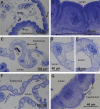
Histological structure of the hindgut epithelium in the anterior chamber and papillate region. A Ventral-lateral epithelium in the anterior chamber B Apical parts of ventral and lateral epithelial cells are bulging into the hindgut lumen C Dorsal epithelium in the anterior chamber forms typhlosole (T) and two typhlosole channels (TC) D Epithelial cell of typhlosole E Epithelial cell of typhlosole channel F Hindgut epithelium in the papillate region G Basal parts of epithelial cells in the papillate region are bulging into haemocoel. Abbreviations: M – muscles, N – cell nucleus.
Figure 3.
Figure 3.
Cell width, cell height and cell nucleus diameter in the two hindgut regions. Scatter plot depicting median values of cell width, cell height and cell nucleus diameter in the anterior chamber against median values of same parameters in the papillate region of seven specimens. The central line is the line of equality. Points lying on the line indicate that the median value of measured parameter is equal in both hindgut regions. Points below the line indicate that the median value of measured parameter is larger in the anterior chamber than in the papillate region. Points above the line indicate that the median value of measured parameter is larger in the papillate region than in the anterior chamber.
Figure 4.
Figure 4.
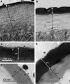
Ultrastructure of the cuticle in the anterior chamber and the papillate region. A The cuticle in the anterior chamber consists of thin epicuticle (EPI) and thick procuticle (PRO) with distinct sublayers. Between the epicuticule and the procuticle, a thin layer of intermediate electron density is present (arrow) B Cuticle in the papillate region consists of epicuticle (EPI) which is proportionally thicker according to the procuticle (PRO). Procuticle sublayers are less pronounced than in the anterior chamber. Between the epicuticle and the procuticle a thin layer of intermediate electron density is present (arrow) C, D The outer part of the epicuticle (EPI) in the anterior chamber (C) and in the papillate region (D) is three-layered (arrow) and covered with a fuzzy layer (arrowhead) E Procuticle (PRO) in the papillate region can be of approximately the same thickness as epicuticle (EPI) and without apparent sublayers.
Figure 5.
Figure 5.
Cuticle and basal lamina thickness in the two hindgut regions. Median values of cuticle thickness (A) and basal lamina thickness (B) in cells from the anterior chamber (blue) and the papillate region (red) of three specimens.
Figure 6.
Figure 6.
Ultrastructure of junctions between cuticle and apical plasma membrane. A Junctions between cuticle and apical plasma membrane in the anterior chamber visible as electron dense structures (arrows), from which fibers (arrowheads) extend into the procuticle B Junctions between cuticle and apical plasma membrane in the papillate region (arrowheads) associated with abundant bundles of microtubules inside the cell (arrow) C, D Higher resolution images of bundles of microtubules (MTB) associated with junctions between cuticle and apical plasma membrane. Image D displays the area denoted on image C (red rectangle) where individual microtubules (arrows) can be discerned.
Figure 7.
Figure 7.
Bacteria associated with hindgut cuticle in the papillate region of one specimen. A bacteria are visible near or at the cuticle (C) surface. Bacterial cells are connected to each other with thin filamentous structures (arrow) B bacteria are connected to the fuzzy layer of epicuticle (arrow) by thin filamentous structures (arrowheads).
Figure 8.
Figure 8.
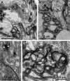
Ultrastructure of the apical plasma membrane labyrinth in the anterior chamber and the papillate region. A Apical membrane labyrinth (AL) in the anterior chamber. In the cytoplasmic strands mitochondria (arrowheads) are present B A tubular appearance of cytoplasmic strands is evident in certain sections (arrow) C Apical membrane labyrinth (AL) in the papillate region. Key: arrowheads – mitochondria, CUT – cuticle.
Figure 9.
Figure 9.
Ultrastructure of the basal plasma membrane labyrinth in the anterior chamber and the papillate region. A Basal plasma membrane in the anterior chamber forms sparse narrow infoldings (arrows) B Extensive basal membrane labyrinth (BL) in the papillate region is associated with numerous mitochondria (arrowheads) C Basal lamina in the anterior chamber. Hemidesmosome-like junctions are visible as small electron dense plaques (arrowheads) D Basal lamina in the papillate region. Hemidesmosome-like junctions are visible as small electron dense plaques (arrowheads). Abbreviation: M – muscle.
Figure 10.
Figure 10.
Apical and basal labyrinth depth and density of membrane infoldings in the two hindgut regions. Scatter plots depict: A Median values of apical labyrinth depth versus the median values of basal labyrinth depth in cells from the anterior chamber (blue) and the papillate region (red) of three specimens B Median values of apical infoldings density against the median values of basal infoldings density in cells from the anterior chamber (blue) and the papillate region (red) of three specimens. The central line in both plots is the line of equality. Points lying on the line indicate that median value of depth/density is equal, apically and basally. Points below the line indicate that the median value of depth/density is larger apically than basally. Points above the line indicate that the median value is larger basally than apically.
Figure 11.
Figure 11.
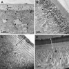
Ultrastructure of cell junctions in the anterior chamber and the papillate region. A The junctional complex in the anterior chamber consists of subapically located adherens junctions (AJ) and septate junctions (SJ) located beneath the adherens junctions B The junctional complex in the papillate region consists of subapically located adherens junctions (AJ) and extremely long and convoluted septate junctions (SJ) located beneath the adherens junctions C Adherens junctions consist of two electron dense plaques (arrowheads) at the cytoplasmic sides of lateral plasma membranes of two neighbouring cells and electron dense material in the intercellular space between the membranes D Septate junctions are visible as electron dense septa arranged in strings (arrow). Dilated intercellular spaces are visible where septate junctions are locally interrupted (arrowhead). Abbreviations: C – cuticle, AI – apical infoldings, SJ – septate junction.
Figure 12.
Figure 12.
Lateral parts of epithelial cells in the anterior chamber and the papillate region. A Two neighbouring cells in the anterior chamber B Two neighbouring cells in the papillate region. Abbreviations: LID – area of lateral interdigitations, AL – apical labyrinth, BL – basal labyrinth, M – muscle.
Figure 13.
Figure 13.
Localization of Na+/K+-ATPase activity in hindguts of intermoult and postmoult animals. A Na+/K+-ATPase activity in the anterior chamber of intermoult P.scaber. Electron dense deposits are present along tubular (arrowheads) and dilated (arrows) apical membranous infoldings and in the procuticle (PRO) B Na+/K+-ATPase activity in the papillate region of postmoult P.scaber. Abundant deposits are present along the apical infoldings (arrowheads) and in the procuticle (PRO) C Control section in the anterior chamber of intermoult animal where K+ ions were replaced by Na+ ions. No reaction product is present along apical infoldings (AI) or in the cuticle (C) D Control section in the papillate region of postmoult animal treated with ouabain. Some deposits (arrowheads) are present along the apical infoldings. Abbreviations: C – cuticle.

References

    1. Berridge MJ, Gupta BL. (1967) Fine-structural changes in relation to ion and water transport in the rectal papillae of the blowfly, Calliphora. Journal of Cell Science 2: 89–112. http://jcs.biologists.org/content/2/1/89 - PubMed
    1. Bettica A, Witkus R, Vernon GM. (1987) Ultrastructure of the foregut-hindgut junction in Porcellioscaber Latreille. Journal of Crustacean Biology 7(4): 619–623. https://www.jstor.org/stable/1548647
    1. Bitsch C, Bitsch J. (2002) The endoskeletal structures in arthropods: cytology, morphology and evolution. Arthropod Structure & Development 30(3): 159–177. 10.1016/S1467-8039(01)00032-9 - DOI - PubMed
    1. Blaxter ML, Page AP, Rudin W, Maizels RM. (1992) Nematode surface coats: Actively evading immunity. Parasitology Today 8(7): 243–247. 10.1016/0169-4758(92)90126-M - DOI - PubMed
    1. Boudour-Boucheker N, Boulo V, Charmantier-Daures M, Grousset E, Anger K, Charmantier G, Lorin-Nebel C. (2014) Differential distribution of V-type H+-ATPase and Na+/K+-ATPase in the branchial chamber of the palaemonid shrimp Macrobrachiumamazonicum. Cell and Tissue Research 357(1): 195–206. 10.1007/s00441-014-1845-5 - DOI - PubMed

LinkOut - more resources