. 2018 Dec 4:9:1668.
doi: 10.3389/fpls.2018.01668. eCollection 2018.

Genome Mining of Plant NPFs Reveals Varying Conservation of Signature Motifs Associated With the Mechanism of Transport

Affiliations

Genome Mining of Plant NPFs Reveals Varying Conservation of Signature Motifs Associated With the Mechanism of Transport

Antonella Longo et al. Front Plant Sci. .

Abstract

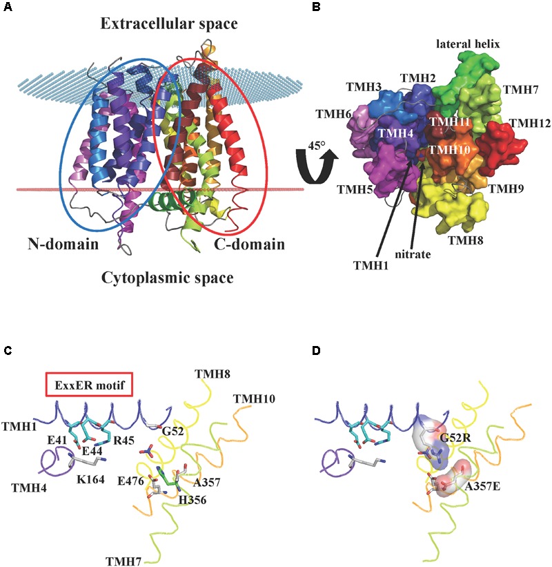
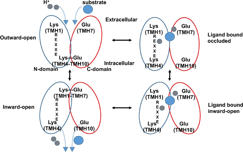
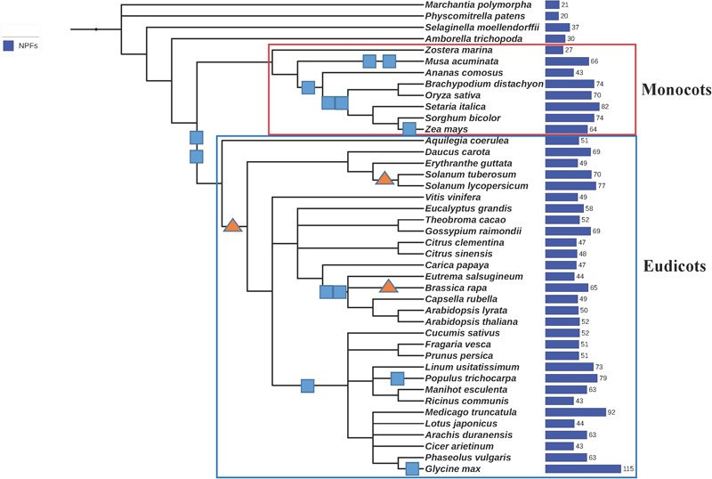
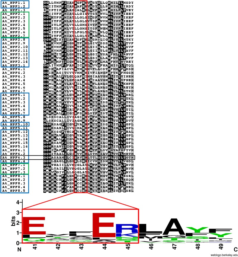
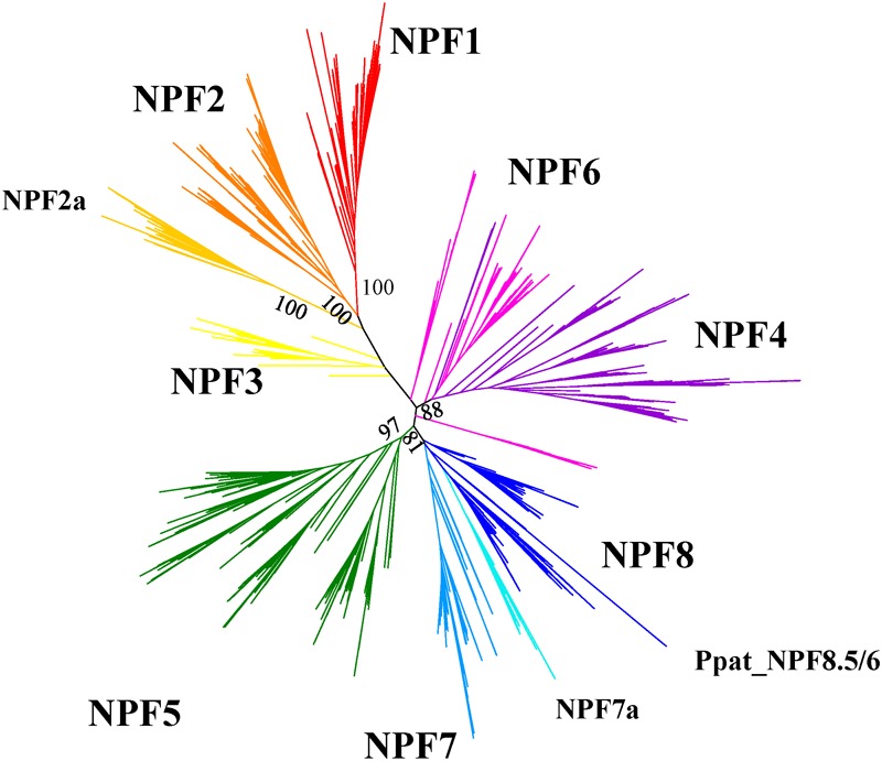
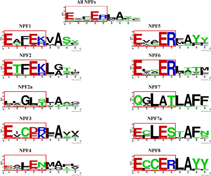
Nitrogen is essential for all living species and may be taken up from the environment in different forms like nitrate or peptides. In plants, members of a transporter family named NPFs transport nitrate and peptides across biological membranes. NPFs are phylogenetically related to a family of peptide transporters (PTRs) or proton-coupled oligopeptide transporters (POTs) that are evolutionarily conserved in all organisms except in Archaea. POTs are present in low numbers in bacteria, algae and animals. NPFs have expanded in plants and evolved to transport a wide range of substrates including phytohormones and glucosinolates. Functional studies have shown that most NPFs, like POTs, operate as symporters with simultaneous inwardly directed movement of protons. Here we focus on four structural features of NPFs/POTs/PTRs that have been shown by structural and functional studies to be essential to proton-coupled symport transport. The first two features are implicated in proton binding and transport: a conserved motif named ExxER/K, located in the first transmembrane helix (TMH1) and a D/E residue in TMH7 that has been observed in some bacterial and algal transporters. The third and fourth features are two inter-helical salt bridges between residues on TMH1 and TMH7 or TMH4 and TMH10. To understand if the mechanism of transport is conserved in NPFs with the expansion to novel substrates, we collected NPFs sequences from 42 plant genomes. Sequence alignment revealed that the ExxER/K motif is not strictly conserved and its conservation level is different in the NPF subfamilies. The proton binding site on TMH7 is missing in all NPFs with the exception of two NPFs from moss. The two moss NPFs also have a positively charged amino acid on TMH1 that can form the salt bridge with the TMH7 negative residue. None of the other NPFs we examined harbor residues that can form the TMH1-TMH7 salt bridge. In contrast, the amino acids required to form the TMH4-TMH10 salt bridge are highly conserved in NPFs, with some exceptions. These results support the need for further biochemical and structural studies of individual NPFs for a better understanding of the transport mechanism in this family of transporters.

Keywords: genome; gibberellin); glucosinolates (GSL); nitrate; nitrate peptide family (NPF) transporters; nitrogen; phytohormones (auxin; proton-dependent oligopeptide transporter.


Figures

FIGURE 1
FIGURE 1
Crystal structure of AtNPF6.3. (A) Cartoon representation of the A. thaliana dual affinity nitrate transporter AtNPF6.3/NRT1.1 (PDB: 4OH3) (Sun et al., 2014) showing the core 12 transmembrane helices (TMHs) arranged into N- and C-terminal bundles (blue and red ovals, respectively). The transporter is in an inward-open conformation with the opening facing the cytoplasm. Spatial position of AtNPF6.3 in the membrane was calculated using the PPM server (Lomize et al., 2012): membrane boundaries are shown as red and blue dots. (B) AtNPF6.3 structure as seen from the cytoplasmic side. Nitrate is located at the bottom of the substrate channel. (C) Close-in view of the substrate channel formed by TMH1, 4, 7, and 10. Important side chains are shown as sticks: Glu41, Glu44 and Arg45 belong to the ExxER/K motif; Lys164 and Glu476 can potentially form an inter-helical salt bridge between TMH4 and TMH10 in the outward open conformation; residues Gly52 and Ala357 correspond to residues that have been shown to form a salt bridge between TMH1 and TMH7 in some bacterial and algal POTs/NRT1s. (D) In silico mutagenesis shows that when residues Gly52 and Ala357 are mutated into arginine and glutamate, respectively, they are within hydrogen bond distance and could form a salt bridge stabilizing the inward-open conformation.
FIGURE 2
FIGURE 2
Proposed alternating-access mechanism in NPF/POT transporters. Schematic model of the proposed NPF/POT alternating-access transport cycle showing four conformations (based on Newstead, 2015). In the outward-open conformation, the transporter is open towards the extracellular space; a salt bridge forms between residues on TMH4 and TMH10, holding the N- and C-terminal bundles (blue and red ovals, respectively) together and stabilizing the conformation. Negatively charged amino acids in TMH7 and in the ExxER/K motif on TMH1 are exposed in the internal cavity and are available for proton binding. After protons (gray spheres) and substrate (blue sphere) bind, the protein undergoes conformational changes that include the disruption of the TMH4–TMH10 salt bridge allowing the transporter to open towards the intracellular space in the inward-open conformation. Finally, protons and substrate are released in the cytoplasm. A salt bridge may form in the inward-open conformation between oppositely charged residues on TMH1 and TMH7 as observed in some crystal structures from bacterial POTs.
FIGURE 3
FIGURE 3
Phylogenetic tree of the 42 plants whose genomes were used in this study. We generated a phylogenetic tree from a list of taxonomic names of the 42 plants analyzed in this study using the online tool phyloT, a phylogenetic tree generator based on NCBI taxonomy (http://phylot.biobyte.de/). The tree was visualized using iTOL (http://itol.embl.de/). Whole genome duplications (WGD) are represented by squares, whole genome triplications are represented by triangles (based on Lee et al., 2012, 2017). The total number of NPFs contained in each genome is reported on the right. Monocots and eudicots are included in the red and blue rectangles, respectively.
FIGURE 4
FIGURE 4
Conservation of the ExxER/K motif in A. thaliana NPFs. Multiple sequence alignment of the TMH1 region of 52 Arabidopsis thaliana NPFs. Included in the sequence are two glycine residues located N-terminal to the TMH1, but not part of the α-helix. The ExxER/K motif (red box) is fully conserved in 31 AtNPFs (60%) (blue box), while ten AtNPFs (19%) completely lack the motif (green box). A logo was created to represent the motif sequence conservation. Amino acid numbering is based on the sequence of AtNPF6.3. Amino acids are colored based on their chemical properties (hydrophobic amino acids are black, polar are green, basic are red, and acidic are blue). The overall height of stacks represents the sequence conservation while the height of letters indicates the relative frequency of each amino acid in that position (Crooks et al., 2004).
FIGURE 5
FIGURE 5
Unrooted maximum-likehood phylogenetic tree of 2383 NPFs sequences from 42 plants visualized with iTOL (Letunic and Bork, 2016). Eight subfamilies are represented with different colors and named based on the nomenclature proposed in Léran et al. (2014). Two subfamilies with specific characteristics regarding the ExxER/K motif, as shown in Figure 6, are labeled as NPF2a and NPF7a. Percent bootstrap values are given for the main branches and support the distribution in subfamilies. The phylogenetic tree can be accessed at https://itol.embl.de/shared/FPS_2018.
FIGURE 6
FIGURE 6
LOGOS of the ExxER/K motif sequence in the NPF subfamilies. Graphical representation of sequence conservation with the transmembrane helix 1 (TMH1) using all NPF sequences or NPF sequences from each subfamily separately. Numbering and coloring as in Figure 4.

References

    1. Almagro A., Lin S. H., Tsay Y. F. (2008). Characterization of the Arabidopsis nitrate transporter NRT1.6 reveals a role of nitrate in early embryo development. Plant Cell 20 3289–3299. 10.1105/tpc.107.056788 - DOI - PMC - PubMed
    1. Arnold K., Bordoli L., Kopp J., Schwede T. (2006). The SWISS-MODEL workspace: a web-based environment for protein structure homology modelling. Bioinformatics 22 195–201. 10.1093/bioinformatics/bti770 - DOI - PubMed
    1. Biasini M., Bienert S., Waterhouse A., Arnold K., Studer G., Schmidt T., et al. (2014). SWISS-MODEL: modelling protein tertiary and quaternary structure using evolutionary information. Nucleic Acids Res. 42 W252–W258. 10.1093/nar/gku340 - DOI - PMC - PubMed
    1. Boggavarapu R., Jeckelmann J. M., Harder D., Ucurum Z., Fotiadis D. (2015). Role of electrostatic interactions for ligand recognition and specificity of peptide transporters. BMC Biol. 6:58. 10.1186/s12915-015-0167-8 - DOI - PMC - PubMed
    1. Capella-Gutiérrez S., Silla-Martínez J. M., Gabaldón T. (2009). trimAl: a tool for automated alignment trimming in large-scale phylogenetic analyses. Bioinformatics 25 1972–1973. 10.1093/bioinformatics/btp348 - DOI - PMC - PubMed

LinkOut - more resources