. 2018 Nov 14;5(11):181166.
doi: 10.1098/rsos.181166. eCollection 2018 Nov.

Short cracks in knee meniscus tissue cause strain concentrations, but do not reduce ultimate stress, in single-cycle uniaxial tension

Affiliations

Short cracks in knee meniscus tissue cause strain concentrations, but do not reduce ultimate stress, in single-cycle uniaxial tension

John M Peloquin et al. R Soc Open Sci. .

Abstract

Tears are central to knee meniscus pathology and, from a mechanical perspective, are crack-like defects (cracks). In many materials, cracks create stress concentrations that cause progressive local rupture and reduce effective strength. It is currently unknown if cracks in meniscus have these consequences; if they do, this would have repercussions for management of meniscus pathology. The objective of this study was to determine if a short crack in meniscus tissue, which mimics a preclinical meniscus tear, (a) causes crack growth and reduces effective strength, (b) creates a near-tip strain concentration and (c) creates unloaded regions on either side of the crack. Specimens with and without cracks were tested in uniaxial tension and compared in terms of macroscopic stress-strain curves and digital image correlation strain fields. The strain fields were used as an indicator of stress concentrations and unloaded regions. Effective strength was found to be insensitive to the presence of a crack (potential effect < 0.86 s.d.; β = 0.2), but significant strain concentrations, which have the potential to lead to long-term accumulation of tissue or cell damage, were observed near the crack tip.

Keywords: crack; fracture mechanics; mechanical failure; meniscus tear; rupture; tensile test.


Conflict of interest statement

We declare we have no competing interests.

Figures

Figure 1.
Figure 1.
Schematic of ultimate stress variation with fracture toughness and stress concentration severity. Fracture mechanics governs failure when the material's fracture toughness is small relative to a local stress concentration. Limit stress analysis governs failure when the material is sufficiently tough to tolerate any stress concentrations that are present. In the fracture mechanics case, σuUTS; in the limit stress case, σu = σUTS [23,24,26].
Figure 2.
Figure 2.
Specimen schematics. (a) Sites of specimen dissection. (b) Specimen shapes for each of the analysis groups (circumferential tension 90° edge crack, circumferential tension 45° centre crack and radial tension 90° centre crack) and their crack-free controls.
Figure 3.
Figure 3.
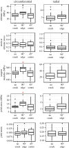
Comparison of stress–strain curve parameters between cracked specimens and their corresponding controls. The boxplots are in Tukey’s style. Asterisks (*) indicate cracked case ≠ control case, Welch t-test, p < 0.05.
Figure 4.
Figure 4.
Stress–strain curves for all analysed specimens.
Figure 5.
Figure 5.
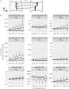
Strain field differences between near-tip and away-from tip ROIs within specimens. (a) Schematic of ROI definitions. (b) Tukey boxplots. Asterisks (*) indicate near ≠ away, Wilcoxon test, p < 0.05.
Figure 6.
Figure 6.
Representative strain fields for cracked specimens in each group at σ = 0.7σu. Scale bar, 5 mm.
Figure 7.
Figure 7.
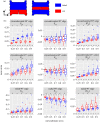
Strain field differences between cut and intact ROIs within specimens. (a) ROI definitions. (b) Tukey boxplots. Asterisks (*) indicate cut ≠ intact, paired Wilcoxon test, p < 0.05.
Figure 8.
Figure 8.
Examples of cracked specimen rupture morphology. (a) Circumferential control specimen, showing a widespread rupture zone and interdigitating fibre pull-out that is typical of circumferential specimen rupture. (b) Circumferential edge crack specimen, showing early rupture at the crack tip combined with crack blunting by inter-fascicle shear. (c) Circumferential centre crack specimen, showing the typical outcome of independent rupture sites merging with the crack. (d) Circumferential centre crack specimen, showing zigzagging of a rupture across the crack and along fascicle interfaces. (e) Radial edge crack specimen, showing crack growth. (f) Radial edge crack specimen, showing simultaneous rupture across its entire width. (af) Scale bar, 5 mm.

References

    1. Englund M, Roemer FW, Hayashi D, Crema MD, Guermazi A. 2012. Meniscus pathology, osteoarthritis and the treatment controversy. Nat. Rev. Rheumatol. 8, 412–419. (10.1038/nrrheum.2012.69) - DOI - PubMed
    1. Øiestad BE, Engebretsen L, Storheim K, Risberg MA. 2009. Knee osteoarthritis after anterior cruciate ligament injury: a systematic review. Am. J. Sports Med. 37, 1434–1443. (10.1177/0363546509338827) - DOI - PubMed
    1. Cohen M, Amaro JT, Ejnisman B, Carvalho RT, Nakano KK, Peccin MS, Teixeira R, Laurino CFS, Abdalla RJ. 2007. Anterior cruciate ligament reconstruction after 10 to 15 years: association between meniscectomy and osteoarthrosis. Arthroscopy 23, 629–634. (10.1016/j.arthro.2007.03.094) - DOI - PubMed
    1. Englund M, Guermazi A, Gale D, Hunter DJ, Aliabadi P, Clancy M, Felson DT. 2008. Incidental meniscal findings on knee MRI in middle-aged and elderly persons. New England J. Med. 359, 1108–1115. (10.1056/NEJMoa0800777) - DOI - PMC - PubMed
    1. Magnussen RA, Mansour AA, Carey JL, Spindler KP. 2009. Meniscus status at anterior cruciate ligament reconstruction associated with radiographic signs of osteoarthritis at 5- to 10-year follow-up: a systematic review. J. Knee Surg. 22, 347–357. (10.1055/s-0030-1247773) - DOI - PMC - PubMed

LinkOut - more resources