

Laboratory and Neuroimaging Biomarkers in Neuropsychiatric Systemic Lupus Erythematosus: Where Do We Stand, Where To Go?
- PMID: 30564579
- PMCID: PMC6288259
- DOI: 10.3389/fmed.2018.00340
Laboratory and Neuroimaging Biomarkers in Neuropsychiatric Systemic Lupus Erythematosus: Where Do We Stand, Where To Go?
Abstract
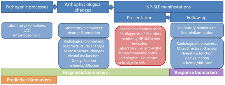
Systemic lupus erythematosus (SLE) is a chronic autoimmune disease characterized by multi-systemic involvement. Nervous system involvement in SLE leads to a series of uncommon and heterogeneous neuropsychiatric (NP) manifestations. Current knowledge on the underlying pathogenic processes and their subsequent pathophysiological changes leading to NP-SLE manifestations is incomplete. Several putative laboratory biomarkers have been proposed as contributors to the genesis of SLE-related nervous system damage. Alongside the laboratory biomarkers, several neuroimaging tools have shown to reflect the nature of tissue microstructural damage associated with SLE, and thus were suggested to contribute to the understanding of the pathophysiological changes and subsequently help in clinical decision making. However, the number of useful biomarkers in NP-SLE in clinical practice is disconcertingly modest. In some cases it is not clear whether the biomarker is truly involved in pathogenesis, or the result of non-specific pathophysiological changes in the nervous system (e.g., neuroinflammation) or whether it is the consequence of a concomitant underlying abnormality related to SLE activity. In order to improve the diagnosis of NP-SLE and provide a better targeted care to these patients, there is still a need to develop and validate a range of biomarkers that reliably capture the different aspects of disease heterogeneity. This article critically reviews the current state of knowledge on laboratory and neuroimaging biomarkers in NP-SLE, discusses the factors that need to be addressed to make these biomarkers suitable for clinical application, and suggests potential future research paths to address important unmet needs in the NP-SLE field.
Keywords: NP-SLE; biomarkers; magnetic resonance imaging; neuroimaging; neuropsychiatric systemic lupus erythematosus; systemic lupus erythematosus.
Figures
References
-
- Magro-Checa C, Zirkzee EJ, Beaart-van de Voorde LJJ, Middelkoop HA, van der Wee NJ, Huisman MV, et al. Value of multidisciplinary reassessment in attribution of neuropsychiatric events to systemic lupus erythematosus: prospective data from the Leiden NPSLE cohort. Rheumatology (2017) 56:1676–83. 10.1093/rheumatology/kex019 - DOI - PubMed
Publication types
LinkOut - more resources
Full Text Sources
Miscellaneous