. 2018 Oct 23;6(2):233-238.
doi: 10.1016/j.ijvsm.2018.10.001. eCollection 2018 Dec.

Ultrasonography guidance for total splenectomy in donkeys

Affiliations

Ultrasonography guidance for total splenectomy in donkeys

Kamal-Eldeen Fouad et al. Int J Vet Sci Med. .

Abstract

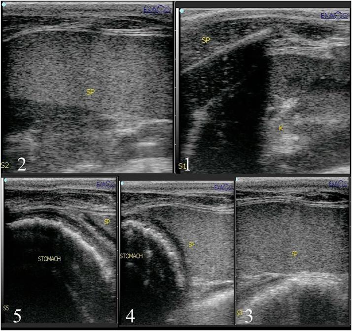
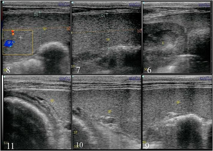
There are varieties of surgical approaches reported for equine splenectomy and all of them were dealing with the most reachable situation of splenic hilus and easy handling of the spleen. The aim of this work was to establish the normal ultrasound parameters of spleen in donkeys (normal echogenicity, hilus situation, topographic location and correlation with neighboring organs) as a guide to select the best approach for total splenectomy in donkeys. Splenic ultrasound was carried out on six normal donkeys before experimental total splenectomy in the standing position. The splenic topographic location was recorded among 4 rows including 30 squares. These animals were divided into two groups according to the surgical approach of total splenectomy. Total splenectomy after left 16th and 17th ribs partial resection in standing position was carried out in group1 and group 2, respectively. Ultrasonographically, the spleen had homogenously echogenic pattern and appeared hyperechoic to the liver. Only one third of the spleen was located in front of the 16th rib where the hilus and splenic blood vessels were nearly under the 16th rib. The splenic artery and splenic vein were ultrasonographically visualized between the left 16th and 17th ribs 10-15 cm from dorsal midline. This area was the site of the important ligation during total splenectomy. In conclusion, ultrasonography guidance for total splenectomy in donkeys assisted the surgical findings and proved that technique following partial resection of the 17th rib at the standing position is the most convenient surgical approach for total splenectomy in donkeys.

Keywords: Intercostal spaces; Rib; Spleen; Splenomegaly; Ultrasound.


Figures

Fig. 1
Fig. 1
Splenectomy in a donkey. (a) Left gastric epiploic, short gastric vessels (yellow arrow) and splenic blood vessels were double ligated. (b) One ligature (black arrow) was laid close to the stomach and the other was close to the spleen. (c) A cut was made between the ligatures. (d) The excised spleen. (For interpretation of the references to colour in this figure legend, the reader is referred to the web version of this article.)
Fig. 2
Fig. 2
(a) Topographical anatomy of the spleen in a donkey. (b) Ultrasongraphic findings of spleen in the scanned area.
Fig. 3
Fig. 3
Ultrasonography at upper part of the left flank region showing left kidney and small part of spleen base (1), U/S between left 18th and 17th ribs showing part of splenic base (2), U/S between left 17th and 16th ribs showing part of splenic base (3), U/S between left 16th and 15th ribs showing small part of splenic base and stomach (4) and U/S between left 15th and 14th ribs showing small part of splenic base and stomach (5).
Fig. 4
Fig. 4
Splenic ultrasonography at the left flank region caudal to left 18th rib showing small part of spleen and left kidney (6), U/S between left 18th and 17th ribs showing part of splenic body (7), U/S between left 17th and 16th ribs showing spleen and splenic artery (8), U/S between left 16th and 15th ribs showing splenic body (9), U/S between left 15th and 14th ribs showing splenic body and stomach (10) and U/S between left 14th and 13th ribs showing small part of splenic body and stomach (11).
Fig. 5
Fig. 5
Ultrasonography in the middle part of left flank region showing intestine (12), U/S at the flank region under left 18th rib showing small part of spleen and intestine (13), U/S between left 17th and 16th ribs showing splenic body (14), U/S between left 16th and 15th ribs showing splenic body (15), U/S between left 17th and 16th ribs showing splenic body (16), U/S between left 14th and 13th ribs showing splenic body (17), U/S between left 13th and 12th ribs showing splenic body (18), U/S between left 12th and 11th ribs showing splenic body (19) and U/S between left 11th and 10th ribs showing splenic body (20).
Fig. 6
Fig. 6
Ultrasonography of the most lower part of left flank region showing intestine (21), (22), (23) and spleen appeared at the level of lower abdomen between left 16th and 15th ribs (24), U/S between left 15th and 14th ribs showing splenic apex (25), U/S between left 14th and 13th ribs showing splenic apex (26), U/S between left 13th and 12th ribs showing splenic apex (27), U/S between left 12th and 11th ribs showing splenic apex (28), U/S between left 11th and 10th ribs showing splenic apex (29) and U/S between left 10th and 9th ribs showing splenic apex (30).

References

    1. Sgorbini M., Bonelli F., Rota A., Baragli P., Marchetti V., Corazza M. Hematology and clinical chemistry in amiata donkey foals from birth to 2 months of age. J Equine Vet Sci. 2012;33:35–39.
    1. Getachew A., Burden F., Wernery U. Common infectious diseases of working donkeys: their epidemiological and zoonotic role. J Equine Vet Sci. 2016;39:S107.
    1. Alsop E.J., Marr C., Barrelet A.B., Mcgladdery A.J. The use of transabdominal ultrasonography in the diagnosis and management of splenic lesions in three horses. Equine Vet Educ. 2007;19:5–10.
    1. Reef V.B. WB Saunders; Philadelphia, PA: 1998. Equine Diagnostic Ultrasound; pp. 273–363.
    1. Freeman S.L. Diagnostic ultrasonography of the mature equine abdomen. Equine Vet Educ. 2003;15:319–330.