. 2018 Oct 30;5(12):1543-1561.
doi: 10.1002/acn3.664. eCollection 2018 Dec.

TLR3 agonism re-establishes CNS immune competence during α 4-integrin deficiency

Affiliations

TLR3 agonism re-establishes CNS immune competence during α 4-integrin deficiency

Rehana Z Hussain et al. Ann Clin Transl Neurol. .

Abstract

Objective: Natalizumab blocks α4-integrin-mediated leukocyte migration into the central nervous system (CNS). It diminishes disease activity in multiple sclerosis (MS), but carries a high risk of progressive multifocal encephalopathy (PML), an opportunistic infection with JV virus that may be prompted by diminished CNS immune surveillance. The initial host response to viral infections entails the synthesis of type I interferons (IFN) upon engagement of TLR3 receptors. We hypothesized that TLR3 agonism reestablishes CNS immune competence in the setting of α4-integrin deficiency.

Method: We generated the conditional knock out mouse strain Mx1.Cre+ α4-integrinfl/fl, in which the α4-integrin gene is ablated upon treatment with the TLR3 agonist poly I:C. Adoptive transfer of purified lymphocytes from poly I:C-treated Mx1.Cre+ α4-integrinfl/fl donors into naive recipients recapitulates immunosuppression under natalizumab. Active experimental autoimmune encephalomyelitis (EAE) in Mx1.Cre+ α4-integrinfl/fl mice treated with poly I:C represents immune-reconstitution.

Results: Adoptive transfer of T cells from poly I:C treated Mx1.Cre+ α4-integrinfl/fl mice causes minimal EAE. The in vitro migratory capability of CD45+ splenocytes from these mice is reduced. In contrast, actively-induced EAE after poly I:C treatment results in full disease susceptibility of Mx1.Cre+ α4-integrinfl/fl mice, and the number and composition of CNS leukocytes is similar to controls. Extravasation of Evans Blue indicates a compromised blood-brain barrier. Poly I:C treatment results in a 2-fold increase in IFN β transcription in the spinal cord.

Interpretation: Our data suggest that TLR3 agonism in the setting of relative α4-integrin deficiency can reestablish CNS immune surveillance in an experimental model. This pathway may present a feasible treatment strategy to treat and prevent PML under natalizumab therapy and should be considered for further experimental evaluation in a controlled setting.


Figures

Figure 1
Figure 1
Generating a model to assess the effects of Toll‐like receptor 3 (TLR3) agonism on CNS immune re‐constitution in the setting of relative α4‐integrin deficiency. We hypothesized that agonism of TLR3 with polyinosinic‐polycytidylic acid (poly I:C) would fully reestablish EAE disease activity in mice that lack α4‐integrin. To address this hypothesis, we generated the Mx1.Cre+ α4‐integrinflfl mouse strain as described in the Methods section. In these mice, the Cre recombinase is under the control of the Mx1 promoter which can be induced to high levels by administration of poly I:C. (A) Poly I:C engagement of TLR3 results in the endosomal compartment (B) leads to the activation of interferon (IFN) regulatory factors (IRF) and nuclear factor kappa‐light‐chain‐enhancer of activated B cells (NF κB) in the cytosol and the transcription factor activator protein‐1 (AP‐1), and eventually in the transcription and expression of type I interferons, which (C) subsequently bind IFN type I receptors in adjacent IFN Type I receptor‐expressing cells. All tissues and organs and all hematopoietic cells express IFN type I receptors.81 Activation of tyrosine kinase 2 (TYK2) and Janus kinase 1 (JAK1) ensues, and (D) downstream transcription factors, including signal transducer and activator of transcription 1 (STAT1) translocate to the cell nucleus, and start transcription of antiviral genes, including Mx1. In Mx1.Cre+ α4‐integrinfl/fl mice, Cre recombinase targets loxP sites flanking the Itga4 (α4‐integrin) gene, causing its deletion. (E) This model allows the conditional deletion of α4‐integrin on all IFN type I receptor‐expressing cells, which includes leukocytes. Adoptive transfer experiments, in vitro migration assays, and some leukocyte immunophenotyping (all Fig. 2) were performed in the absence of poly I:C, and therefore only represent the immunosuppressive aspects of this model.
Figure 2
Figure 2
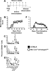
In vivo and in vitro Characterization of Mx1.Cre+ α4‐integrinfl/fl mice with or without exposure of the Toll‐like receptor 3 (TLR3) agonist polyinosinic‐polycytidylic acid (poly I:C). (A) To ablate α4‐integrin, Mx1.Cre + α4 fl/fl mice received three intra peritoneal injections of 300 μg poly(I)‐poly(C) (poly I:C; Sigma Chemical Company) given at 2 days intervals in order to activate the Cre recombinase. This was followed by a “wash‐out” period of three weeks in which mice were then analyzed or immunized for EAE. (B) EAE disease incidence, onset, clinical severity are similar between Mx1.Cre+ α4‐integrinfl/fl mice and C57BL/6 control mice not exposed to poly I:C. Then, the frequency of α4‐integrin (CD49d)‐expressing leukocyte subsets was assessed by multi‐parameter flow cytometry in poly I:C‐treated Mx1.Cre+ α4‐integrinfl/fl mice, and in poly I:C‐treated C57BL/6 control mice on day 15 post active EAE induction. (C) In the lymph nodes, (D) spleen, and (E) bone marrow of poly I:C‐treated Mx1.Cre+ α4‐integrinfl/fl mice, the frequency of α4‐integrin expressing CD3+ T cells, CD8+ T cells, CD11c+ monocyte‐derived dendritic cells (DC), and CD22b+Ly6G+ myeloid‐derived granulocytes is significantly diminished. (D) In spleen, and (E) bone marrow, the frequency of α4‐integrin expressing CD4+ T cells, CD19+ B cells, and CD22b+Ly6G macrophages is also significantly reduced. (C) Similar trends for the latter cell subsets are seen in lymph nodes but do not reach statistical significance. Next, we tested the in vivo role of genetic α4‐integrin ablation by passively transferring activated cells from myelin oligodendrocyte glycoprotein peptide (MOG p) 35‐55‐immunized poly I:C‐treated Mx1.Cre+ α4‐integrinfl/fl mice, or poly I:C‐treated C57BL/6 mice into naïve C57BL/6 recipient mice. In the adoptive transfer model, the recipient mice are not exposed to the effects of poly I:C. (F) Transfer of cells from poly I:C‐treated Mx1.Cre+ α4‐integrinfl/fl donor T cells was associated with decreased disease and severity in recipient mice.
Figure 3
Figure 3
Lymph node cells and splenocytes from systemically poly I:C‐treated Mx1.Cre+ α4‐integrinfl/fl show altered migratory behavior in vitro. To test the effect of α4‐integrin deletion after poly I:C treatment on migratory competence of cells, we performed an in vitro migration assay on lymph node cells and splenocytes by Boyden Chamber as described before.27 Briefly, a total of 5 × 10⁵ splenocytes from mice actively immunized for EAE and sacrificed at day 10, suspended in media, were added to the upper chamber. Chambers were then incubated at 37°C for 6 h. Following incubation, the content of the lower chamber was collected, and the number of cells was counted with a hemocytometer and the phenotype of the cells determined by flow cytometry. (A) The composition of lymph node cells from poly I:C treated C57BL/6 control mice and (B) poly I:C‐treated Mx1.Cre+ α4‐integrinfl/fl mice entered into the upper chamber, and (C and D) the composition of lymph node cells of both mouse strains that migrated into the lower chamber were assessed by multi‐parameter flow cytometry. (A and B) There were subtle differences in the composition of lymph node cells between the mouse strains at the outset of the migration experiments. After 6 h of migration, the composition in the lower chamber reflected (C) an enhanced migratory capability of conventional CD11c+ dendritic cells (DC) in poly I:C‐treated Mx1.Cre+ α4‐integrinfl/fl mice, and (D) a reduced migratory capability of Ly6G+ neutrophils compared to poly I:C treated C57BL/6 control mice. (E) Overall, there was a non‐significant reduction in the migration of CD45+ lymph node cells of poly I:C‐treated Mx1.Cre+ α4‐integrinfl/fl mice, which was driven by diminished migration of (F) CD4+ T cells, CD8+ T cells, CD19+ B cells, and (G) Ly6G+ neutrophils. (H) The composition of splenocytes from poly I:C treated C57BL/6 control mice and (I) poly I:C‐treated Mx1.Cre+ α4‐integrinfl/fl mice in the upper chamber showed a decreased percentage of Ly6G+ neutrophils in Mx1.Cre+ α4‐integrinfl/fl mice, which was compensated by an expansion of all other leukocytes subsets. (J and K) The composition of splenocytes in the lower chamber was similar between mouse strains. (L) In splenocytes, there was also a non‐significant reduction in the migration of CD45+ lymph node cells of poly I:C‐treated Mx1.Cre+ α4‐integrinfl/fl mice, which was driven by diminished migration of (M) CD19+ B cells, and (N) Ly6G+ neutrophils.
Figure 4
Figure 4
In vivo systemic Toll‐like receptor 3 (TLR3) agonism through polyinosinic‐polycytidylic acid (poly I:C) treatment reestablishes EAE disease susceptibility and CNS immune competence in the setting of relative α4‐integrin deficiency. (A) When active EAE was induced in Mx1.Cre+ α4‐integrinfl/fl mice and C57BL/6 control mice that were treated with poly I:C, EAE disease incidence, susceptibility, and severity were similar in both groups. Next, the percent of leukocytes in (B) lymph nodes, (C) spleen, (D) brain, and (E) spinal cord nodes was assessed by multi‐parameter flow cytometry in mice that were actively immunized for EAE and treated with poly I:C on disease day 15. There were no differences in composition of leukocytes between the two strains, indicating a full cellular immune re‐constitution. (F) Utilizing the CellTrace™ CFSE (5(6)‐carboxyfluorescein N‐hydroxysuccinimidyl ester) Cell Proliferation kit (Life Technologies, Carlsbad, CA), we detected no difference between the capacity of Mx1.Cre+ α4‐integrinfl/fl mice and C57BL/6 control mice that were treated with poly I:C to mount recall responses to MOG p35‐55. (G) The number of activated CD4+ CD25+ T cells was increased in the brain of Mx1.Cre+ α4‐integrinfl/fl mice treated with poly I:C, and similar between both mouse strains in the spinal cord. (H) In the brain and spinal cord, we also observed a significant expansion of CD19+ SSC hi B cells in Mx1.Cre+ α4‐integrinfl/fl mice treated with poly I:C.
Figure 5
Figure 5
Toll‐like receptor 3 (TLR3) agonism through polyinosinic‐polycytidylic acid (poly I:C) compromises the blood‐brain barrier. To test the effect of in vivo poly I:C treatment on blood‐brain barrier BBB integrity, we performed an Evans Blue Dye (EBD) permeability assay. EBD has a high affinity for serum albumin. In the setting of BBB compromise, the serum‐dye complex can penetrate the CNS parenchyma, and it can be visualized and quantified by spectrophotometry. (A) There was no difference in the amount of EBD detected in the CNS of Mx1.Cre+ α4‐integrinfl/fl mice and C57BL/6 control mice treated in vivo with poly I:C. (B–E) We also did not observe a difference in the absolute number of inflammatory infiltrates in the spinal cords between animals of both mouse strains in whom active EAE had been induced in the absence or presence of poly I:C. The anatomical locations of BBB compromise as indicated by EBD extravasation differed in the (F and G) brain, and (H and I) spinal cord differed between Mx1.Cre+ α4‐integrinfl/fl mice and C57BL/6 control mice treated in vivo with poly I:C.
Figure 6
Figure 6
In vivo Toll‐like receptor 3 (TLR3) agonism through systemic polycytidylic acid (poly I:C) promotes diverse integrin usage in CNS‐infiltrating. To determine the integrin usage required for leukocytes migration into the brain and spinal cords, we assessed the expression of (A–D) Lymphocyte‐function associated antigen‐1 (LFA‐1; β2‐integrin; CD11a), (E–H) α5‐integrin (CD49e), and (I–L) α4‐integrin (CD49d) on different lymphocyte and myeloid cell subsets in Mx1.Cre+ α4‐integrinfl/fl mice and C57BL/6 control mice actively induced for EAE on day 15 by multi‐parameter flow cytometry. (A–L) The number of leukocyte subsets expressing CD11a, CD49e, and CD49d in all compartments was similar between mouse strains.
Figure 7
Figure 7
Systemic Toll‐like receptor 3 (TLR3) agonism through polycytidylic acid (poly I:C) differentially impacts cytokine expression in a compartment‐specific manner in the setting of relative α4‐integrin deficiency. (A) Engagement of TLR3 results in the transcription and cellular expression of the type I interferon beta (IFN β) in spinal cord. To confirm that systemic administration of poly I:C induces transcription of type I IFN within the CNS, we performed quantitative real time polymerase chain reation (qPCR) for IFN β in the brain and spinal cord of Mx1.Cre+ α4‐integrinfl/fl mice 24 h after administration of three doses of poly I:C on consecutive days. In the spinal cord, there was an approximate 2‐fold increase in IFN β transcripts compared to tissue from untreated mice. (B) On day 15 after active induction of experimental autoimmune encephalomyelitis (EAE), or 36 days after the last dose of poly I:C, we observed a significant increase in the transcription of interleukin (IL)‐1 beta (β), and a substantial increase in the transcription of IL‐1α, IL‐6, IL‐12, IL‐17, granulocyte‐macrophage colony‐stimulating factor (GMCSF), and interferon gamma (IFN γ) compared to mice not treated with poly I:C in the spinal cord.

Similar articles

Cited by

References

    1. Stuve O, Zamvil SS. Neurologic diseases In: Parslow TG, Stites DP, Terr AI, Imboden JB, eds. Medical Immunology. San Francisco: McGraw Hill, 2001:510–526.
    1. Yednock TA, Cannon C, Fritz LC, et al. Prevention of experimental autoimmune encephalomyelitis by antibodies against alpha 4 beta 1 integrin. Nature 1992;356:63–66. - PubMed
    1. Theien BE, Vanderlugt CL, Eagar TN, et al. Discordant effects of anti‐VLA‐4 treatment before and after onset of relapsing experimental autoimmune encephalomyelitis. J Clin Invest 2001;107:995–1006. - PMC - PubMed
    1. Stuve O, Marra CM, Jerome KR, et al. Immune surveillance in multiple sclerosis patients treated with natalizumab. Ann Neurol 2006;59:743–747. - PubMed
    1. Stuve O, Marra CM, Bar‐Or A, et al. Altered CD4+/CD8+ T‐cell ratios in cerebrospinal fluid of natalizumab‐treated patients with multiple sclerosis. Arch Neurol 2006;63:1383–1387. - PubMed