. 2018 Dec 12:2:28.
doi: 10.1038/s41698-018-0071-0. eCollection 2018.

Deep sequencing of circulating exosomal microRNA allows non-invasive glioblastoma diagnosis

Affiliations

Deep sequencing of circulating exosomal microRNA allows non-invasive glioblastoma diagnosis

Saeideh Ebrahimkhani et al. NPJ Precis Oncol. .

Abstract

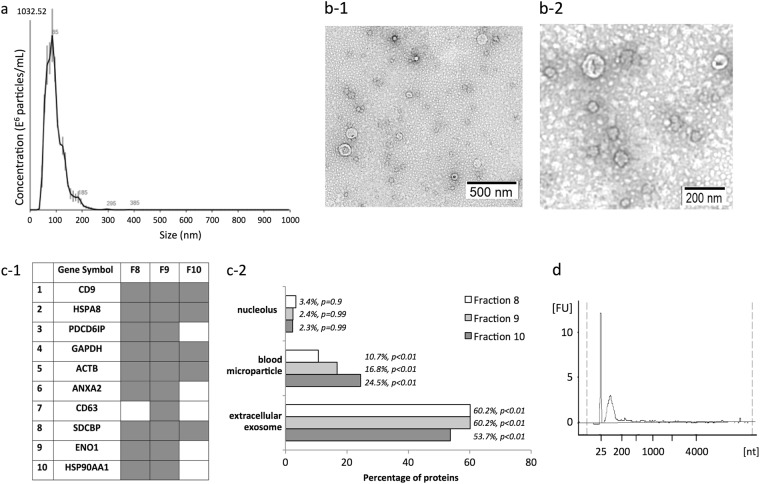
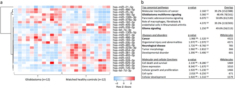
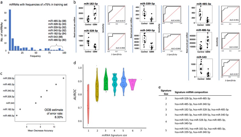
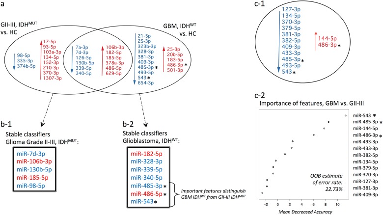
Exosomes are nano-sized extracellular vesicles released by many cells that contain molecules characteristic of their cell of origin, including microRNA. Exosomes released by glioblastoma cross the blood-brain barrier into the peripheral circulation and carry molecular cargo distinct to that of "free-circulating" miRNA. In this pilot study, serum exosomal microRNAs were isolated from glioblastoma (n = 12) patients and analyzed using unbiased deep sequencing. Results were compared to sera from age- and gender-matched healthy controls and to grade II-III (n = 10) glioma patients. Significant differentially expressed microRNAs were identified, and the predictive power of individual and subsets of microRNAs were tested using univariate and multivariate analyses. Additional sera from glioblastoma patients (n = 4) and independent sets of healthy (n = 9) and non-glioma (n = 10) controls were used to further test the specificity and predictive power of this unique exosomal microRNA signature. Twenty-six microRNAs were differentially expressed in serum exosomes from glioblastoma patients relative to healthy controls. Random forest modeling and data partitioning selected seven miRNAs (miR-182-5p, miR-328-3p, miR-339-5p, miR-340-5p, miR-485-3p, miR-486-5p, and miR-543) as the most stable for classifying glioblastoma. Strikingly, within this model, six iterations of these miRNA classifiers could distinguish glioblastoma patients from controls with perfect accuracy. The seven miRNA panel was able to correctly classify all specimens in validation cohorts (n = 23). Also identified were 23 dysregulated miRNAs in IDHMUT gliomas, a partially overlapping yet distinct signature of lower-grade glioma. Serum exosomal miRNA signatures can accurately diagnose glioblastoma preoperatively. miRNA signatures identified are distinct from previously reported "free-circulating" miRNA studies in GBM patients and appear to be superior.


Conflict of interest statement

The authors declare no competing interests.

Figures

Fig. 1
Fig. 1
Characterization of serum exosomes isolated in fractions 8–10 by size exclusion chromatography prior to miRNA sequencing. a Size distribution of particles as analyzed by nanoparticle tracking analysis. b Transmission electron microscopy allowed visualization of vesicles with sizes ranging from 60 to 110 nm in diameter, scale bars = 500 nm (b-1, wide field) and 200 nm (b-2, close-up). c-1 Mass spectrometry-based proteome analysis of size chromatographic elution fractions 8–10 identified all top 10 exosome marker proteins and c-2 showed significant enrichment of proteins characteristic of exosomes and blood microparticles. Proteins identified in fractions 8–10 showed limited, non-significant associations with compartments like the nucleolus, where certain miRNA species are concentrated. d Bioanalyzer trace of RNA extracted from serum exosomes shows the main population of small RNA and no ribosomal RNA
Fig. 2
Fig. 2
a Hierarchical clustering of 26 differentially expressed miRNAs shows clear separation of glioblastoma (GBM) patients and healthy control (HC) exosomal profiles (fold change ≥2 or ≤0.5; unadjusted p values ≤0.05 in all three statistical tests). b Functional pathway analysis of mRNAs targeted by 44 significantly changing miRNA (unadjusted p values ≤0.05 in all three statistical tests) in GBM circulating exosomes. Top canonical pathways, diseases and disorders and molecular and cellular functions are listed with the numbers of overlapping molecules and significance of associations (right-tailed Fisher exact test, p value)
Fig. 3
Fig. 3
a miRNAs appearing in >75 of the 100 partitions (70% training set, 30% test set) were selected as the most stable miRNA classifiers by Random Forest modeling (frequencies are specified in brackets). b Box-and-whisker plots and receiver operator characteristic curves with area under the curve (AUROC) calculations demonstrate the individual discriminatory power of the seven most stable miRNA classifiers. c miRNAs were ordered by the importance of their contribution to discriminating GBM from [healthy] controls; overall out-of-the-bag (OOB) error rate of the seven features was 8.33%. d AUROC measures of all possible combinations of the seven miRNAs previously identified to be the most stable predictors, stratified by the number of miRNAs (signature size) and their distributions, and displayed as violin plots. e miRNA signatures that discriminate between GBM and healthy controls with perfect accuracy
Fig. 4
Fig. 4
a A Venn diagram summarizes the differentially expressed miRNAs between IDHMUT glioma tumor grades II–III (GII–III; n = 10), IDHWT glioblastoma (GBM; n = 12), and corresponding age- and gender-matched healthy controls (HC; fold change ≥2 or ≤0.5; unadjusted p values ≤0.05 in all three statistics tests, i.e., Exact, t test, and Wilcoxon), with 12 overlapping differentially expressed miRNAs. Decreased expression is indicated in blue and increased expression in red. The most stable miRNAs for classifying b-1 GII-III IDHMUT and b-2 GBM IDHWT from HCs are listed and show distinct features. c-1 Summary of differentially expressed miRNAs between the GBM IDHWT and GII-III IDHMUT cohorts and c-2 plot of “importance” of each individual miRNA for discriminating GBM from GII–III; out-of-the-bag (OOB) error rate is 22.73%. Three of the top four features that distinguish GBM IDHWT from GII–III IDHMUT were only identified in the GBM vs HC comparative analysis, are members of the GBM miRNA signature that together accurately classify GBMs from HCs, and may be specific markers for GBM (indicated by asterisks in a, b-2, c-1, c-2)

Comment in

References

    1. Ellingson BM, Wen PY, Cloughesy TF. Modified criteria for radiographic response assessment in glioblastoma clinical trials. Neurotherapeutics. 2017;14:307–320. doi: 10.1007/s13311-016-0507-6. - DOI - PMC - PubMed
    1. Saadatpour L, et al. Glioblastoma: exosome and microRNA as novel diagnosis biomarkers. Cancer Gene Ther. 2016;23:415–418. doi: 10.1038/cgt.2016.48. - DOI - PubMed
    1. Skog J, et al. Glioblastoma microvesicles transport RNA and proteins that promote tumour growth and provide diagnostic biomarkers. Nat. Cell Biol. 2008;10:1470–1476. doi: 10.1038/ncb1800. - DOI - PMC - PubMed
    1. Mallawaaratchy DM, et al. CompreheGlioblastoma Biomarker withnsive proteome profiling of glioblastoma-derived extracellular vesicles identifies markers for more aggressive disease. J. Neurooncol. 2017;131:233–244. doi: 10.1007/s11060-016-2298-3. - DOI - PMC - PubMed
    1. Hallal, S. et al. Extracellular Vesicles from Neurosurgical Aspirates Identifies Chaperonin Containing TCP1 Subunit 6A (CCT6A) as aPotential Glioblastoma Biomarker with Prognostic Significance. Proteomics 0, 1800157, 10.1002/pmic.201800157 (2018). - PubMed