. 2019 Mar;68(3):421-432.
doi: 10.1007/s00262-018-2282-1. Epub 2018 Dec 18.

A secreted PD-L1 splice variant that covalently dimerizes and mediates immunosuppression

Affiliations

A secreted PD-L1 splice variant that covalently dimerizes and mediates immunosuppression

Kathleen M Mahoney et al. Cancer Immunol Immunother. 2019 Mar.

Abstract

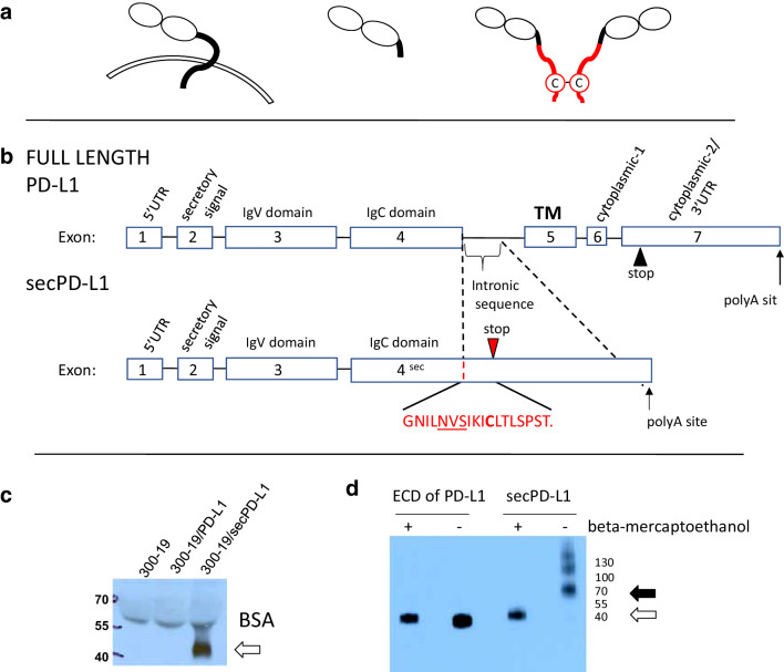
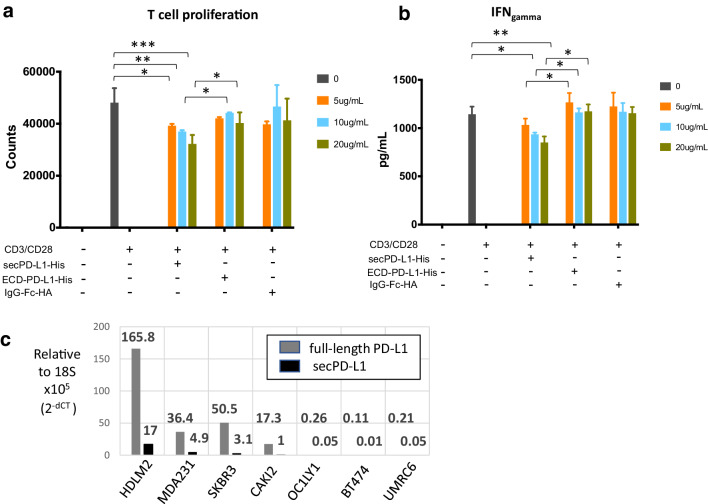
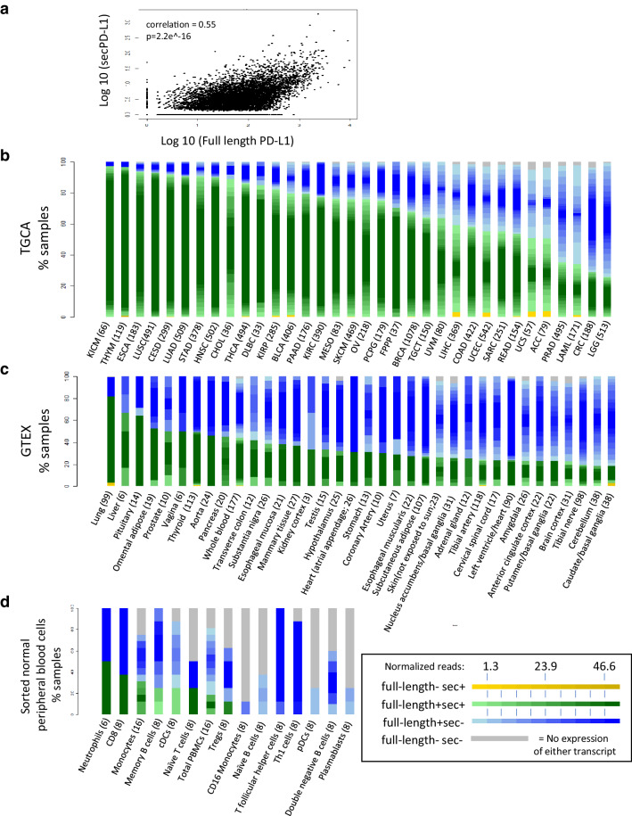
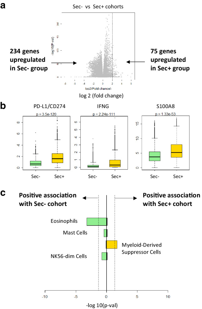
Targeting immune checkpoint pathways, such as programmed death ligand-1 (PD-L1, also known as CD274 or B7-H1) or its receptor programmed cell death-1 (PD-1) has shown improved survival for patients with numerous types of cancers, not limited to lung cancer, melanoma, renal cell carcinoma, and Hodgkin lymphoma. PD-L1 is a co-inhibitory molecule whose expression on the surface of tumor cells is associated with worse prognosis in many tumors. Here we describe a splice variant (secPD-L1) that does not splice into the transmembrane domain, but instead produces a secreted form of PD-L1 that has a unique 18 amino acid tail containing a cysteine that allows it to homodimerize and more effectively inhibit lymphocyte function than monomeric soluble PD-L1. We show that recombinant secPD-L1 can dimerize and inhibit T-cell proliferation and IFN-gamma production in vitro. The secPD-L1 variant is expressed by malignant cells in vitro that also express high levels of full-length PD-L1. Transcriptomic analysis of gene expression across The Cancer Genome Atlas found the strongest association of secPD-L1 with full-length PD-L1, but also with subsets of immunologic genes, such as in myeloid-derived suppressor cells. Moreover, the splice variant is also expressed in normal tissues and within normal peripheral blood cells it is preferentially expressed in activated myeloid cells. This is the first report of a form of secreted PD-L1 that homodimerizes and is functionally active. SecPD-L1 may function as a paracrine negative immune regulator within the tumor, since secPD-L1 does not require a cell-to-cell interaction to mediate its inhibitory effect.

Keywords: Immune checkpoint; Isoforms; PD-L1; Splice variants.


Conflict of interest statement

Shukla has equity in 152 Therapeutics. Freeman and Boussiotis have patents/pending royalties on the PD-1 pathway from Bristol-Myers-Squibb, Roche, Merck, EMD-Serono, Boehringer-Ingelheim, AstraZeneca, Dako, and Novartis. Freeman has a patent application for the use of 9A11 antibody for diagnostic purposes. Hacohen and Wu are founders of Neon Therapeutics and members of its scientific advisory board. Patent applications have been filed: Compositions and Methods for Personalized Neoplasia Vaccines (Hacohen, Fritsch, and Wu), Methods for Identifying Tumor Specific Neo-Antigens (Hacohen and Wu), and Combination Therapy for Neoantigen Vaccine (Hacohen, Wu, and Fritsch). Fritsch is a co-founder and employee of Neon Therapeutics, Inc. All other authors declare that they have no conflict of interest.

Figures

Fig. 1
Fig. 1
A splice variant of PD-L1 mRNA encodes a soluble form of PD-L1 that can covalently dimerize. a Protein schema of transmembrane PD-L1 (left) and a soluble PD-L1 cleaved from the cell surface (middle) and the dimeric secPD-L1 splice variant (right, C indicates a cysteine in the carboxyl-terminal domain). b Exons and introns of full-length PD-L1 (top) and secPD-L1 splice variant with unique cysteine-containing carboxyl-terminal domain (bottom; N-linked glycosylation motif (NVS) underlined). c Western blot analysis of PD-L1 in cell-free supernatants from 300.19 cells expressing either full-length PD-L1 or secPD-L1 (open arrow). d Western blot analysis of recombinant extracellular domain of PD-L1 and secPD-L1, in reducing and non-reducing conditions (open arrow indicating monomeric PD-L1 and closed arrow indicating dimeric secPD-L1 protein)
Fig. 2
Fig. 2
SecPD-L1 is functional and is expressed in PD-L1 “positive” tumor cells. a Proliferation of CD3/CD28 activated T lymphocytes in the presence of recombinant secPD-L1, the soluble monomeric extracellular domain of PD-L1 (ECD-PD-L1) or an immunoglobulin-HA tagged fusion protein as control after 72 h (*p value < 0.05, **p value < 0.005, ***p value < 0.0005). b IFN-γ production of CD3/CD28-activated T lymphocytes after treatment as in a was assayed after 48 h. c Quantitative RT-PCR of full-length and secPD-L1 in tumor cell lines (lymphoma: HDLM2, OC1-LY1; kidney cancer: CAKI-2, UMRC6; and breast cancer: SKBR3, MDA231 and BT474)
Fig. 3
Fig. 3
SecPD-L1 RNA expression is associated with full-length PD-L1 expression and is found in the majority of human tumors and also some normal hematologic cells. a Correlation of secPD-L1 expression with full-length PD-L1 RNA expression in The Cancer Genome Atlas (TCGA) data (normalized to total RNA transcripts, n = 9677 tumors, correlation = 0.55, p = 2.2e− 16). b Frequency of secPD-L1 and/or full-length PD-L1 within each tumor type in the TCGA. Each specimen was assigned to one of four classes: full-length− sec+ (yellow shades), full-length + sec+ (green shades), full-length+ sec− (blue shades), full-length− sec− (grey) based on the frequency of the secPD-L1 and full-length PD-L1 isoforms (number of specimens per tumor type listed in parenthesis). The expression value of a sample within any class was color-shaded as follows. The interquartile range of the overall distribution of both isoforms was divided into 10 equal groups, with the first and last groups subsuming values ≤ 25th and ≥ 75th quartile, respectively. The expression of a sample within a class was represented by the appropriate color on the class-specific color gradient (color gradient created using colorRampPalette in R; full-length− sec+: yellow–yellow, full-length + sec+: light green–dark green, full-length+ sec−: light blue–blue, full-length− sec−: grey). The level of full-length PD-L1 counts is represented by the gradient of color of the full-length+ sec+ and full-length+ sec− groups; the level of secPD-L1 count is represented by the color yellow gradient in the full-length− sec+ group. c Frequency of secPD-L1 and/or full-length PD-L1 in normal tissue in GTEx (number of specimens per tissue type in parenthesis). The expression of the full-length+ sec+ and full-length+ sec− classes was represented by the normalized full-length PD-L1 count while the full-length− sec+ group was represented by the corresponding normalized secPD-L1 count, with darker color representing higher expression of full-length PD-L1 or secPD-L1, respectively. d Some lymphocytes, myeloid cells and neutrophils expressing full-length PD-L1 also express secPD-L1 (secPD-L1+ and full-length PD-L1+ in green, secPD-L1 negative and full-length PD-L1+ in blue, and secPD-L1 negative and full-length PD-L1 negative in grey; transcripts normalized to total transcripts)
Fig. 4
Fig. 4
Gene Set Enrichment Analysis (GSEA) shows differences in sec-expressing and no-sec-expressing cohorts of TCGA. a Differential gene expression of the sec-expressing cohort (75 genes, FDR 5%, greater than twofold change in expression) and no-sec-expressing cohort (234 genes). b Within the 805 immune related genes in the differential gene analysis, PD-L1(CD274), IFNg, and S100A8 were the three most significantly enriched in sec-expressing cohort. c Zlatko’s gene sets overlap analysis indicates that the MDSC gene set is associated with sec + cohort (yellow)

References

    1. Mahoney KM, et al. A secreted PD-L1 splice variant expressed across tumor types inhibits lymphocyte function. J Immunother Cancer. 2016;4(Suppl 1):82.
    1. Mahoney KM et al (2018) A secreted PD-L1 splice variant that covalently dimerizes and mediates immunosuppression. Kidney Cancer Symposium, November 3, 2018, Miami, Florida (oral presentation) - PMC - PubMed
    1. Freeman GJ, et al. Engagement of the PD-1 immunoinhibitory receptor by a novel B7 family member leads to negative regulation of lymphocyte activation. J Exp Med. 2000;192(7):1027–1034. doi: 10.1084/jem.192.7.1027. - DOI - PMC - PubMed
    1. Thompson RH, et al. Tumor B7-H1 is associated with poor prognosis in renal cell carcinoma patients with long-term follow-up. Cancer Res. 2006;66(7):3381–3385. doi: 10.1158/0008-5472.CAN-05-4303. - DOI - PubMed
    1. Finkelmeier F, et al. High levels of the soluble programmed death-ligand (sPD-L1) identify hepatocellular carcinoma patients with a poor prognosis. Eur J Cancer. 2016;59:152–159. doi: 10.1016/j.ejca.2016.03.002. - DOI - PubMed

LinkOut - more resources