. 2019 Mar;137(3):467-485.
doi: 10.1007/s00401-018-1950-8. Epub 2018 Dec 18.

Circulating AQP4-specific auto-antibodies alone can induce neuromyelitis optica spectrum disorder in the rat

Affiliations

Circulating AQP4-specific auto-antibodies alone can induce neuromyelitis optica spectrum disorder in the rat

Sophie Hillebrand et al. Acta Neuropathol. 2019 Mar.

Abstract

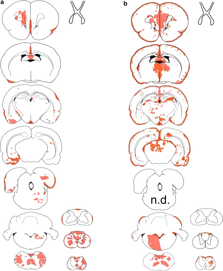
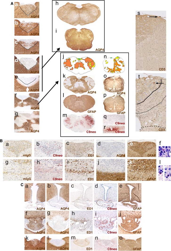
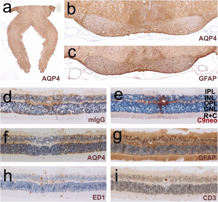
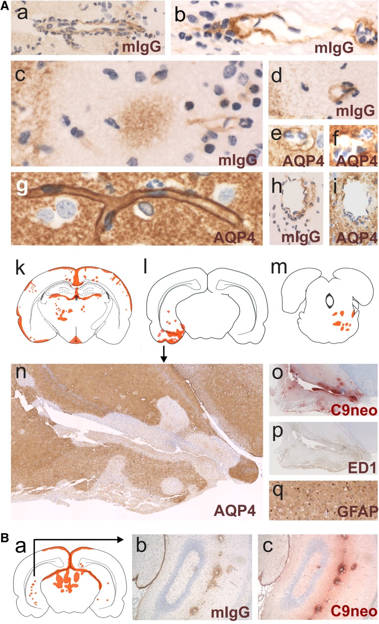
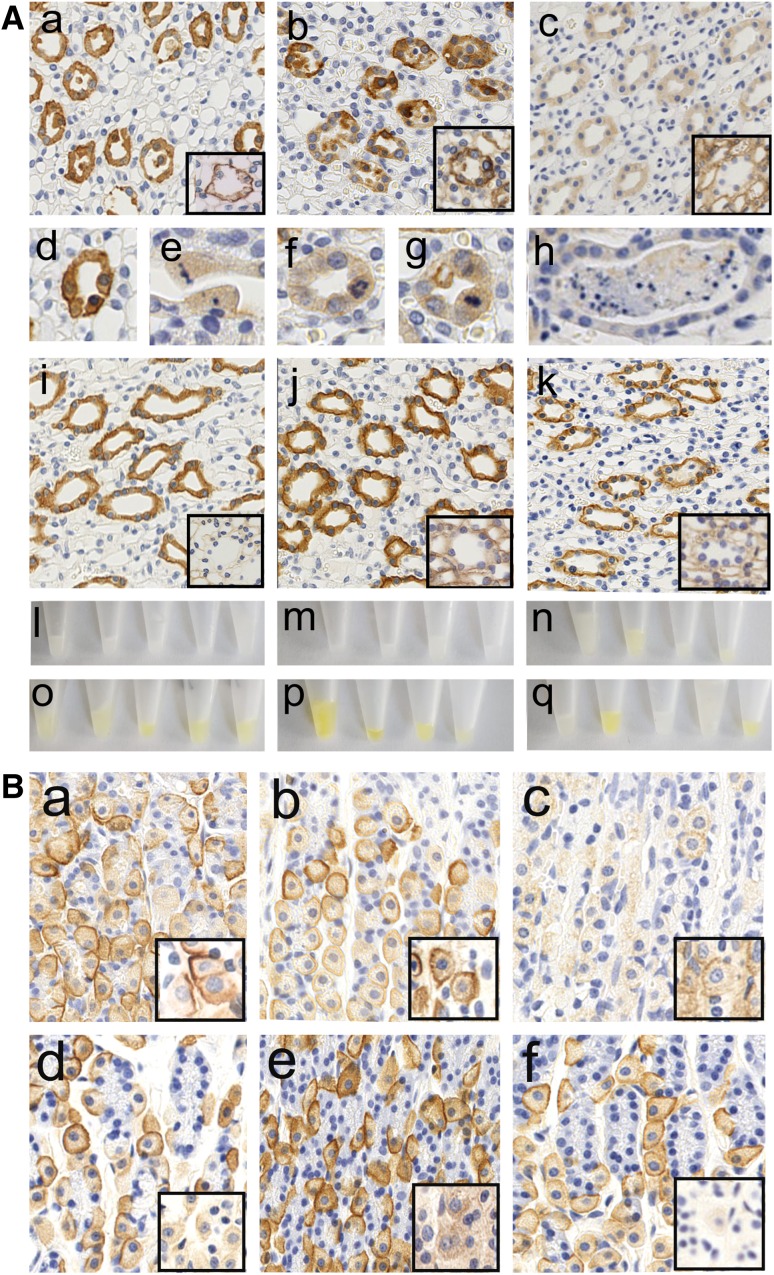
It is well established that the binding of pathogenic aquaporin-4 (AQP4)-specific autoantibodies to astrocytes may initiate a cascade of events culminating in the destruction of these cells and in the formation of large tissue-destructive lesions typical for patients with neuromyelitis optica spectrum disorders (NMOSD). To date, not a single experimental study has shown that the systemic presence of the antibody alone can induce any damage to the central nervous system (CNS), while pathological studies on brains of NMOSD patients suggested that there might be ways for antibody entry and subsequent tissue damage. Here, we systemically applied a highly pathogenic, monoclonal antibody with high affinity to AQP4 over prolonged period of time to rats, and show that AQP4-abs can enter the CNS on their own, via circumventricular organs and meningeal or parenchymal blood vessels, that these antibodies initiate the formation of radically different lesions with AQP4 loss, depending on their mode and site of entry, and that lesion formation is much more efficient in the presence of encephalitogenic T-cell responses. We further demonstrate that the established tissue-destructive lesions trigger the formation of additional lesions by short and far reaching effects on blood vessels and their branches, and that AQP4-abs have profound effects on the AQP4 expression in peripheral tissues which counter-act possible titer loss by antibody absorption outside the CNS. Cumulatively, these data indicate that directly induced pathological changes caused by AQP4-abs inside and outside the CNS are efficient drivers of disease evolution in seropositive organisms.

Keywords: Aquaporin-4; Aquaporin-4-specific antibodies; Kidney; Lesions; Neuromyelitis optica; T cells.


Conflict of interest statement

SH, KS, MN, IT, DB, PP, YT, IN, TM, and MB: the authors declare that they have no conflict of interest. HL received honoraria for lectures from Novartis, Biogen, and Sanofi Aventis. Moreover, he is a member of advisory boards at Roche and Medday. The University Hospital and Medical University of Innsbruck (Austria, MR) receives payments for antibody assays (MOG, AQP4, and other autoantibodies) and for MOG and AQP4 antibody validation experiments organized by Euroimmun (Lübeck, Germany). K. Fujihara serves on scientific advisory boards for Bayer, Biogen, Mitsubishi Tanabe, Novartis, Chugai, Ono, Nihon, Merck Serono, Alexion, and MedImmune; has received funding for travel and speaker honoraria from Bayer, Biogen, Eisai, Mitsubishi Tanabe, Novartis, Astellas, Takeda, Asahi Kasei Medical, Daiichi-Sankyo, and Nihon; serves as an editorial board member of Clinical and Experimental Neuroimmunology, Frontiers in Neurology (section Multiple Sclerosis and Neuroimmunology) and Neurology: Neuroimmunology and Neuroinflammation, and an advisory board member of Sri Lanka Journal of Neurology; has received research support from Bayer, Biogen, Asahi Kasei Medical, The Chemo-Sero-Therapeutic Research Institute, Teva, Mitsubishi Tanabe, Teijin, Chugai, Ono, Nihon, and Genzyme; is funded by Grants-in-Aid for Scientific Research from the Ministry of Education, Culture, Sports, Science and Technology of Japan and by Grants-in-Aid for Scientific Research from the Ministry of Health, Welfare and Labor of Japan.

Figures

Fig. 1
Fig. 1
Lesions with aquaporin 4 loss in AQP4-specific antibody-injected rats. Distribution of established lesions with AQP4 loss along the neuraxis, using schemes provided by Paxinos and Watson [33] as guide lines. Shown here are brain and spinal cord [cervical (C1–7), thoracal (T1–10) and lumbar/sacral (L1–S4)] sections, as well as outlines of optic nerve, chiasm, and optic tract of Lewis (a, n = 5) and RNU rats (b, n = 5). The animals were analyzed 120 h after daily intraperitoneal injections of AQP4-abs, and the location of each lesion with AQP4 loss was projected in red color into the relevant scheme
Fig. 2
Fig. 2
Loss of aquaporin 4-reactivity in circumventricular organs. A Time course of AQP4 loss from the area postrema of Lewis rats. AQP4 stainings (brown) of representative area postremae from uninjected animals (Aa), animals injected daily with mouse control IgG and sacrificed after 120 h (Ab), and animals injected daily with the monoclonal AQP4-specific murine E5415A IgG and sacrificed after 24 (Ac), 48 (Ad), and 120 h (Ae–Ag). All sections were counterstained with hematoxylin to reveal nuclei in blue. The dotted black line depicts the outline of the area postremae. Ah, Ai In some cases, AQP4 loss at the area postrema may be the only pathological changes observed in the medulla of Lewis rats and human patients. AQP-4 stained cross sections of the medulla of a Lewis rat injected daily with the monoclonal AQP4-specific murine E5415A IgG and sacrificed after 120 h (Ah) and a patient with NMOSD (Ai). Aj–Ar In other cases, the medulla shows AQP4 loss at the area postrema and additionally also perivascular loss of AQP4 reactivity. Medullary cross sections at the level of the area postrema were made from a Lewis rat injected daily with the monoclonal AQP4-specific murine E5415A antibody and sacrificed after 120 h (Aj–Am), and from an NMOSD patient (An–Ar). The lesions observed were projected into the corresponding schemes (Aj, An), with the area postrema shown in green, lesions with loss of AQP4 reactivity in yellow, and areas with loss of GFAP reactivity in orange. The corresponding stainings with α-AQP4 antibodies (brown, loss of AQP reactivity white; Ak, Ao), with α-GFAP antibodies (dark brown, loss of reactivity pale brown; Al, Ap), and α-complement C9neo (red; Am, Aq, Ar) are shown. Counterstaining was done with hematoxylin to reveal nuclei (blue). Note that, in the experimental animal and in the human NMOSD case, the area postrema does not show complement C9neo reactivity (Am, Aq), while perivascular lesions in distance to the area postrema show complement deposition (Am, Ar). T cells are essentially absent from the medullas of experimental Lewis rats injected with the monoclonal AQP4-specific murine E5415A antibody and sacrificed after 48 (As) and 120 h (At). Shown are medullary sections at the level of the area postrema, stained with α-CD3 antibodies (brown). B Consecutive histological sections of the medulla of an AQP4-specific antibody-injected Lewis rats, 120 h after the initiation of antibody injection. Shown here are the consequences of AQP4 loss for the area postrema (Ba–Bf) and the area subpostrema (Bg–Bl). The sections were stained with antibodies specific for murine IgG (Ba, Bg), complement C9neo (Bb, Bh), ED1 (Bc, Bi), AQP4 (Bd, Bj), and GFAP (Be, Bk). With exception of complement C9neo, where a positive reaction product is shown in red (Bb, Bh), all other antibody reaction products are shown in brown. Counterstaining was done with hematoxylin to reveal nuclei (blue). Eosinophils were visualized by Giemsa staining (Bf, Bl; red). C Loss of aquaporin 4-reactivity in circumventricular organs and parenchymal lesions with aquaporin 4-loss spreading from these sites. Histological sections of eminentia mediana (Ca–Ce), subfornical organ (Cf–Cj), and area postrema (Ck–Co) were stained for AQP4 (brown, Ca, Cb, Cf, Cg, Ck, Cl), ED1 (brown, Cc, Ch, Cm), complement C9neo (red, Cd, Ci, Cn), and GFAP (brown, Ce, Cj, Co). The sections shown derived from control Lewis rats (Ca, Cf, Ck) and from Lewis rats injected daily with the monoclonal AQP4-specific murine E5415A IgG and sacrificed after 120 h. In all sections, counterstaining was done with hematoxylin to reveal nuclei (blue)
Fig. 3
Fig. 3
Subpial loss of aquaporin 4-reactivity. A Ongoing formation of subpial lesions. Shown first is the distribution of established lesions with AQP4 loss in brain and spinal cord of single Lewis rats (Aa, Ah) which had been injected daily with the monoclonal AQP4-specific murine E5415A IgG and sacrificed after 120 h. The location of each established lesion with AQP4 loss was projected in red color into the relevant scheme. The site of the earliest ongoing lesions used for further characterization is indicated by black arrows in CNS. Consecutive sections of these earliest ongoing lesions were then reacted with antibodies against murine IgG (Ab, Ai, and Ao, brown) or against complement C9neo (Ac, Aj, Ap, red), with the ED1 antibody (Ad, Ak, Aq, brown), and antibodies against AQP4 (Ae, Al, Ar, brown), against GFAP (Af, Am, As, brown), and against CD3 (Ag, An, At, brown; please note that, in this picture, parts of the section were folded back). In all sections, counterstaining was done with hematoxylin to reveal nuclei (blue). B Characterization of subpial lesion from an RNU rat injected daily with the monoclonal AQP4-specific murine E5415A IgG and sacrificed after 120 h. Ba Distribution of established lesions with AQP4 loss in this animal is shown along the neuraxis, using schemes provided by Paxinos and Watson [33] as guide lines. Shown here are brain and spinal cord [cervical (C1–7), thoracal (T1–10), and lumbar/sacral (L1–S4)] sections, as well as outlines of optic nerve, chiasm, and optic tract, and the location of each lesion with AQP4 loss was projected in red color into the relevant scheme. The arrow shows the location of the subpial lesion further characterized by stainings with antibodies against murine IgG (Bb, brown, inlay shows the presence of neutrophils), AQP4 (Bc, brown), with the ED1 antibody (Bd, brown), and with antibodies against GFAP (Be, brown), complement C9neo (Bf, red) and CD3 (Bg, brown; please note the complete absence of T cells). In all sections, counterstaining was done with hematoxylin to reveal nuclei (blue). C Characterization of an extensive periventricular lesion formed in an RNU rat which had been injected daily with the monoclonal AQP4-specific murine E5415A IgG and sacrificed after 120 h. Consecutive sections at the level of the third ventricle were reacted with anti-mouse IgG (Ca, brown), anti-AQP4 (Cb, brown), ED1 (Cc, brown), anti-complement C9neo (Cd, red), and anti-CD3 (Ce, brown). In all sections, counterstaining was done with hematoxylin to reveal nuclei (blue)
Fig. 4
Fig. 4
Perivascular loss of aquaporin 4-reactivity. A Localization and immunohistochemical characterization of leaky vessels allowing antibody entry into the CNS parenchyma. Shown here is the lesion load of individual animals, projected in red onto brain and spinal cord schemes provided by Paxinos and Watson [33] as guide lines (Aa, Ah, Am). The ends of the black arrows indicate the exact location of the vessels reacted in consecutive sections with anti-mouse IgG (Ab, Ai, An, brown), anti-complement C9neo (Ac, Ao, red), ED1 (Ad, Aj, Ap, brown), anti-AQP4 (Ae, Ak, Aq, brown), anti-GFAP (Af, Ar, brown), and anti-CD3 (Ag, Al, as, brown). In all sections, counterstaining was done with hematoxylin to reveal nuclei (blue). B Histological characterization of perivascular lesions found in the brain (Ba, Bc, Be) and spinal cord (Bb, Bd, Bf) of Lewis rats injected daily with the monoclonal AQP4-specific murine E5415A IgG and sacrificed after 120 h. Consecutive sections were stained with anti-AQP4 (Ba, Bb; brown), ED1 (Bc, Bd; brown), and anti-complement C9neo (Be, Bf; red). Counterstaining was done with hematoxylin to reveal nuclei (blue). C Demonstration of neutrophils in and of a near-complete absence of T cells from perivascular lesions. Consecutive lesions derived from spinal cord (Ca, Cb, Ce) and brain (Cc, Cd, Cf) of Lewis rats injected daily with the monoclonal AQP4-specific murine E5415A IgG and sacrificed after 120 h, and were reacted with antibodies against AQP4 (Ca, Cc, Cg, Ch) and against CD3 (Cb, Cd, Ce, Cf). The squares with dotted lines in Ca and Cc outline areas shown in higher magnification in Cg and Ch, respectively, to demonstrate the presence of neutrophils. The arrows in cb and cd point to CD3-positive T cells, and the squares with straight lines shown in Cb and Cd indicate lesion areas shown in higher magnification in Ce and Cf, respectively, to show single T cells
Fig. 5
Fig. 5
Histological characterization of chiasm, optic nerves, and retina. Lewis rats were injected daily with the monoclonal AQP4-specific murine E5415A antibody and were sacrificed after 120 h (ai). The sections were reacted with antibodies against AQP4 (a, b, f), GFAP (c, g), murine IgG (d), complement C9neo (e), with the antibody ED1 (h), or with antibodies against CD3 (i). Positive reaction products are shown in brown (ad, fi) and red (e). All sections were counterstained with hematoxylin to reveal nuclei in blue. IPL inner plexiform layer, INL inner nuclear layer, OPL outer plexiform layer, ONL outer nuclear layer, R + C layer of rods and cones
Fig. 6
Fig. 6
Mechanisms of lesion evolution. A Lesions associated with subpial loss of AQP4 reactivity. Shown here are brain sections with blood vessels entering from the meninges, stained with antibodies against murine IgG (Aa–Ad, Ah) or against AQP4 (Ae–Ag, Ai). Positive reaction products are shown in brown. Counterstaining was done with hematoxylin to reveal nuclei (blue). Ak–Am Examples for lesion clusters seen in the brains of individual animals. The lesions were projected in red onto schemes provided by Paxinos and Watson [33] as guide lines. The lesion cluster pointed out by arrow in Al was further reacted with antibodies against AQP4 (An), complement C9neo (Ao), with the antibody ED1 (Ap), or with antibodies against GFAP (Aq). Positive reaction products of complement C9neo are shown in red, all others in brown. Counterstaining was done with hematoxylin to reveal nuclei (blue). B Example for lesions deriving from branches of the longitudinal hippocampal vein. Shown here are the lesions of a single animal, projected in red onto a scheme provided by Paxinos and Watson [33] as guideline. The lesion cluster pointed out by arrow (Ba) was further reacted with antibodies against murine IgG (Bb), and complement C9neo (Bc)
Fig. 7
Fig. 7
Activated, CNS antigen-specific T cells accelerate lesion formation in the presence of the monoclonal AQP4-specific murine E5415A antibody. Spinal cord (af) and brain (g) sections of Lewis rats daily injected with monoclonal AQP4-abs and sacrified 24 h (a, c, g) or 48 h (e) after the initiation of antibody injection (e), and of Lewis rats which were seropositive for monoclonal AQP4-abs injected once in a concentration of 1 mg (b, d, f) or 0.5 mg (g) at the onset of CNS inflammation induced by the activated MBP-specific T cells (b, f) or AQP4268–285-specific T cells (d), and sacrificed 24 h (b, d, g) or 48 h (f) later. The sections were stained with commercial α-AQP4 antibodies to reveal the expression of AQP4 (brown) and counterstained with hematoxylin to reveal nuclei (blue)
Fig. 8
Fig. 8
Loss of AQP4 reactivity from peripheral organs. A Kidney sections of Lewis rats which had been injected daily with the monoclonal AQP4-specific murine E5415A IgG (Aa–Ah) or with murine control IgG (Aj–Ak) were analyzed. Tissue was sampled after 0 h (Ai), 24 h (Aa, Ad, Ae), 48 h (Ab, Af, Ag, Aj), and 120 h (Ac, Ah, Ak), and stained with commercial α-AQP4 antibodies to reveal the expression of AQP4 on the cell membrane of kidney collecting duct epithelial cells (brown, large pictures), or stained with antibodies against murine IgG to reveal the binding of the injected murine E5415A antibody to AQP4 on kidney collecting duct epithelial cells, and the lack of binding of the injected murine control IgG (brown, small inserts with black rims) to these sites. Please note that, after 24–48 h presence of E5415A, detached epithelial cells are found within the lumen of the collecting ducts. Ad–Ah detailed pictures of kidney collecting duct epithelial cells stained with commercial α-AQP4 specific antibodies. Ad–Ae When E5415A was present for 24 h, the epithelial cells show the strong basolateral expression of AQP4, the formation of pycnotic nuclei and ongoing detachment of epithelial cells from the basement membrane. Af–Ag When E5415A was present for 48 h, many epithelial cells show reduced levels of AQP4 expression. The tubular cells are plump and show mitotic figures as evidence for ongoing regeneration. Ah When E5415A was present for 120 h, rare casts of cellular debris were seen in the lumen of collecting ducts. Urine from Lewis rats injected daily with the monoclonal AQP4-specific murine E5415A IgG was isolated after 24 (Al), 48 (Am), or 120 h (An) and compared to urine of uninjected rats (Ao), and to urine of rats daily injected with murine control IgG and collected after 48 (Ap) or 120 h (Aq). B Stomach sections of Lewis rats which had been injected daily with the monoclonal AQP4-specific murine E5415A IgG (Ba, Bc) or with murine control IgG (Bd–Bf). Tissue was sampled after 0 h (Bd), 24 h (Ba), 48 h (Bb, Be), and 120 h (Bc, Bf), and stained with commercial α-AQP4 antibodies to reveal the expression of AQP4 on the cell membrane of parietal cells (brown, large pictures), or stained with antibodies against murine IgG to reveal the binding of the injected murine E5415A antibody to AQP4 on parietal cells, and the lack of binding of the injected murine control IgG (brown, small inserts) to these sites
Fig. 9
Fig. 9
Four possible scenarios of AQP4 antibody-induced tissue damage. a Entry through the circumventricular organs. Here, there is just some protein leakage in the absence of major vascular damage. Thus, moderate levels of autoantibodies will induce AQP4 loss, but there is little complement leakage and little recruitment of effector cells, and the astrocytes survive. b Leakage through meningeal vessels. Since meningeal vessels are not covered by astrocytic foot processes, AQP4 antibody leakage will not result in amplified damage of the blood–brain barrier. Thus, complement activation will be minor in the subpial areas and needs longer to build up. In addition, complement factors leaking from meningeal vessels will be diluted by CSF and washed away by CSF flow. c Leakage through parenchymal vessels. Only by this route, AQP4 antibodies cause massive focal damage to the blood–brain barrier, associated with profound leakage of complement components, which are not washed away due to the narrow extracellular space of the brain parenchyma. d When there is a primary T-cell-mediated inflammation, inflammation will be targeted to the meninges and the parenchymal vessels (not present in circumventricular organs). In this case, inflammation induces damage to the blood–brain barrier, thus allowing entry of AQP4-abs, complement proteins, and effector cells, activates effector cells, and also induces local production of complement components, in particular in macrophages. Thus, only rather low concentrations of specific antibodies are necessary to cause damage to the AQP4 expressing astrocytes by ADCC and CDC

References

    1. Argaw AT, Asp L, Zhang J, Navrazhina K, Pham T, Mariani JN, et al. Astrocyte-derived VEGF-A drives blood–brain barrier disruption in CNS inflammatory disease. J Clin Investig. 2012;122:2454–2468. doi: 10.1172/JCI60842. - DOI - PMC - PubMed
    1. Bennett JL, Lam C, Kalluri SR, Saikali P, Bautista K, Dupree C, et al. Intrathecal pathogenic anti-aquaporin-4 antibodies in early neuromyelitis optica. Ann Neurol. 2009;66:617–629. doi: 10.1002/ana.21802. - DOI - PMC - PubMed
    1. Bradl M, Kanamori Y, Nakashima I, Misu T, Fujihara K, Lassmann H, et al. Pain in neuromyelitis optica-prevalence, pathogenesis and therapy. Nat Rev Neurol. 2014;10:529–5236. doi: 10.1038/nrneurol.2014.129. - DOI - PubMed
    1. Bradl M, Misu T, Takahashi T, Watanabe M, Mader S, Reindl M, et al. Neuromyelitis optica: pathogenicity of patient immunoglobulin in vivo. Ann Neurol. 2009;66:630–643. doi: 10.1002/ana.21837. - DOI - PubMed
    1. Brooks CG, Webb PJ, Robins RA, Robinson G, Baldwin RW, Festing MF. Studies on the immunobiology of rnu/rnu “nude” rats with congenital aplasia of the thymus. Eur J Immunol. 1980;10:58–65. doi: 10.1002/eji.1830100112. - DOI - PubMed

Publication types

LinkOut - more resources