

Short-Term Assessment of Intravitreal Dexamethasone Implant Using Enhanced-Depth Image Optical Coherence Tomography and Optical Coherence Tomography Angiography in Patients with Retinal Vascular Diseases
- PMID: 30565180
- PMCID: PMC6824342
- DOI: 10.1007/s12325-018-0848-0
Short-Term Assessment of Intravitreal Dexamethasone Implant Using Enhanced-Depth Image Optical Coherence Tomography and Optical Coherence Tomography Angiography in Patients with Retinal Vascular Diseases
Abstract
Introduction: To evaluate the short-term efficacy and safety of intravitreal dexamethasone implant (IDI) in patients with macular oedema associated with diabetic retinopathy (DR) and retinal vein occlusion (RVO) using enhanced-depth image optical coherence tomography (EDI-OCT) and to estimate the effect of dexamethasone on the choroid and the retinal vascular network using OCT angiography (OCTA).
Methods: Fifteen eyes in 15 patients with macular oedema secondary to diabetes (DR, n = 8) or retinal vein occlusion (RVO, n = 7) were treated with intravitreal injection of sustained-release IDI. Primary efficacy end points were changes in best corrected visual acuity and central macular thickness (CMT). Secondary end points were changes in choroidal thickness and choroidal and retinal vascular networks as determined by OCTA.
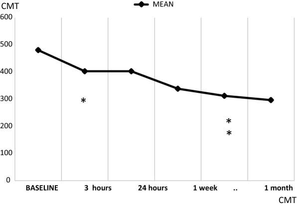
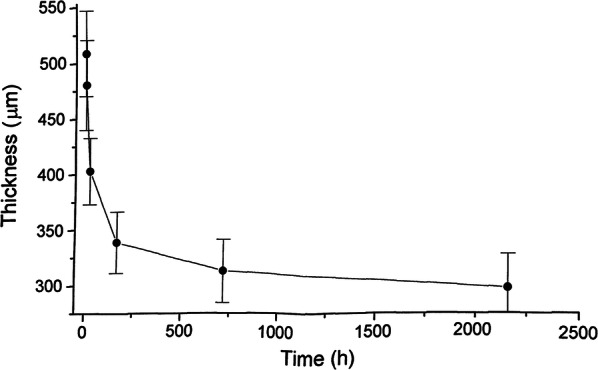
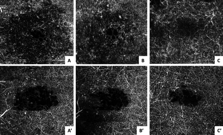
Results: CMT was significantly reduced from baseline by 3 h after injection (p < 0.01) and improved further during the 3-month follow-up. Visual acuity improvement was consistent with CMT reduction. No alterations in IOP or systemic side effects were observed. OCTA showed improvement from baseline in terms of decreased number and size of cysts and restoration of the retinal vascular network; flow choroidal thickness did not change significantly. CMT and visual acuity variations were similar in the two groups.
Conclusions: CMT reduced as early as 3 h after the injection of IDI, with further reduction during follow-up. Choroidal thickness was unchanged, whereas the vascular retinal network improved from baseline to the end of study. Both EDI-OCT and OCTA were useful in demonstrating the early beneficial effects of IDI on the macula and the perifoveal vascular network.
Funding: The article processing charges, the open access fee and the medical writing and editorial assistance was funded by Allergan.
Keywords: Central retinal thickness; Diabetic macular oedema; Enhanced-depth image optical coherence tomography; Innovative biotechnology; Macular oedema; OCT angiography; Ophthalmology; Retinal vein occlusion; Spectral domain OCT; Sustained release intravitreal dexamethasone implant.
Conflict of interest statement
Angelo Maria Minnella, Matteo Federici, Valeria Pagliei, Angela Lanza, Gloria Gambini, Carmela Grazia Caputo, Benedetto Falsini, and Aldo Caporossi have nothing to disclose.
Figures


Similar articles
-
SD-OCT pattern of retinal venous occlusion with cystoid macular edema treated with Ozurdex®.Eur J Ophthalmol. 2011 Sep-Oct;21(5):631-6. doi: 10.5301/EJO.2011.7428. Eur J Ophthalmol. 2011. PMID: 21500185
-
Optical coherence tomography angiography in retinal vein occlusion treated with dexamethasone implant: a new test for follow-up evaluation.Eur J Ophthalmol. 2016 Aug 4;26(5):460-8. doi: 10.5301/ejo.5000829. Epub 2016 Jul 12. Eur J Ophthalmol. 2016. PMID: 27405288
-
Two or more dexamethasone intravitreal implants in treatment-naïve patients with macular edema due to retinal vein occlusion: subgroup analysis of a retrospective chart review study.BMC Ophthalmol. 2015 Sep 4;15:118. doi: 10.1186/s12886-015-0106-z. BMC Ophthalmol. 2015. PMID: 26337664 Free PMC article.
-
Efficacy and safety of dexamethasone versus intravitreal aflibercept implants for macular edema: a systematic review and meta-analysis.Eur J Med Res. 2025 Apr 15;30(1):273. doi: 10.1186/s40001-025-02404-x. Eur J Med Res. 2025. PMID: 40229845 Free PMC article.
-
OZURDEX® (Dexamethasone Intravitreal Implant) 0.7 mg as Initial Therapy in Pseudophakic Patients With Diabetic Macular Edema, Macular Edema Following Retinal Vein Occlusion, and Noninfectious Posterior Segment Uveitis: A Case-Based Discussion.Retina. 2019 Jun;39 Suppl 1:S1-S28. doi: 10.1097/01.iae.0000574292.81105.b2. Retina. 2019. PMID: 31206423 Review. No abstract available.
Cited by
-
Effectiveness of Mp-3 Microperimetric Biofeedback Fixation Training For Low Vision Rehabilitation in Patients Treated With Corticosteroid Ivt in Retinal Vein Occlusions.Clin Optom (Auckl). 2024 May 22;16:131-142. doi: 10.2147/OPTO.S460999. eCollection 2024. Clin Optom (Auckl). 2024. PMID: 38798785 Free PMC article.
-
OCT-Angiography Changes in Patients with Diabetic Macular Edema Treated with Intravitreal Dexamethasone Implant.Clin Ophthalmol. 2022 Feb 2;16:247-263. doi: 10.2147/OPTH.S345947. eCollection 2022. Clin Ophthalmol. 2022. PMID: 35140455 Free PMC article.
-
Short-term effect of intravitreal dexamethasone implant in refractory diabetic macular edema.BMC Ophthalmol. 2024 Mar 11;24(1):113. doi: 10.1186/s12886-024-03341-9. BMC Ophthalmol. 2024. PMID: 38462613 Free PMC article.
References
Publication types
MeSH terms
Substances
Associated data
LinkOut - more resources
Full Text Sources
Medical
