

Vascular Risk and β-Amyloid Are Synergistically Associated with Cortical Tau
- PMID: 30565287
- PMCID: PMC6351182
- DOI: 10.1002/ana.25399
Vascular Risk and β-Amyloid Are Synergistically Associated with Cortical Tau
Abstract
Objective: Neuropathological studies have demonstrated that cerebrovascular disease and Alzheimer disease (AD) pathology frequently co-occur in older adults. The extent to which cerebrovascular disease influences the progression of AD pathology remains unclear. Leveraging newly available positron emission tomography (PET) imaging, we examined whether a well-validated measure of systemic vascular risk and β-amyloid (Aβ) burden have an interactive association with regional tau burden.
Methods: Vascular risk was quantified at baseline in 152 clinically normal older adults (mean age = 73.5 ± 6.1 years) with the office-based Framingham Heart Study cardiovascular disease risk algorithm (FHS-CVD). We acquired Aβ (11 C-Pittsburgh compound B) and tau (18 F-flortaucipir) PET imaging on the same participants. Aβ PET was performed at baseline; tau PET was acquired on average 2.98 ± 1.1 years later. Tau was measured in the entorhinal cortex (EC), an early site of tau deposition, and in the inferior temporal cortex (ITC), an early site of neocortical tau accumulation associated with AD. Linear regression models examined FHS-CVD and Aβ as interactive predictors of tau deposition, adjusting for age, sex, APOE ε4 status, and the time interval between baseline and the tau PET scan.
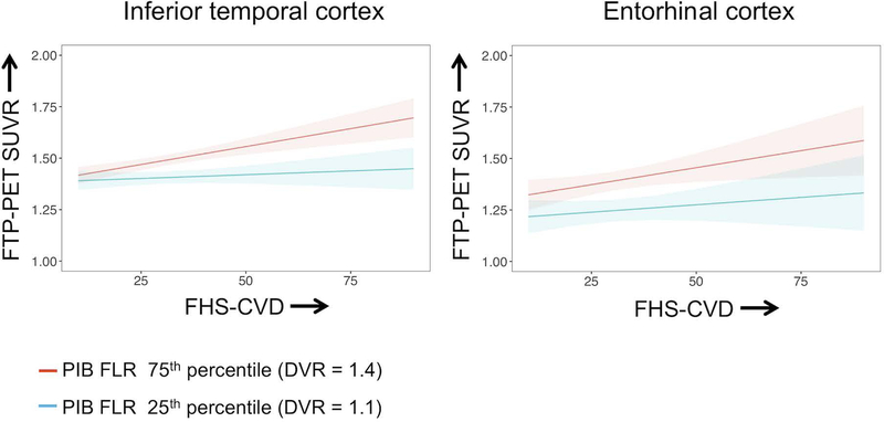
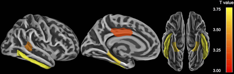
Results: We observed a significant interaction between FHS-CVD and Aβ burden on subsequently measured ITC tau (p < 0.001), whereby combined higher FHS-CVD and elevated Aβ burden was associated with increased tau. The interaction was not significant for EC tau (p = 0.16).
Interpretation: Elevated vascular risk may influence tau burden when coupled with high Aβ burden. These results suggest a potential link between vascular risk and tau pathology in preclinical AD. Ann Neurol 2019; 1-8 ANN NEUROL 2019;85:272-279.
© 2018 American Neurological Association.
Figures
Similar articles
-
Tau Mediates Synergistic Influence of Vascular Risk and Aβ on Cognitive Decline.Ann Neurol. 2022 Nov;92(5):745-755. doi: 10.1002/ana.26460. Epub 2022 Aug 13. Ann Neurol. 2022. PMID: 35880989 Free PMC article.
-
Region-Specific Association of Subjective Cognitive Decline With Tauopathy Independent of Global β-Amyloid Burden.JAMA Neurol. 2017 Dec 1;74(12):1455-1463. doi: 10.1001/jamaneurol.2017.2216. JAMA Neurol. 2017. PMID: 28973551 Free PMC article.
-
Sex Differences in the Association of Global Amyloid and Regional Tau Deposition Measured by Positron Emission Tomography in Clinically Normal Older Adults.JAMA Neurol. 2019 May 1;76(5):542-551. doi: 10.1001/jamaneurol.2018.4693. JAMA Neurol. 2019. PMID: 30715078 Free PMC article.
-
Cerebral small vessel disease and the risk of Alzheimer's disease: A systematic review.Ageing Res Rev. 2018 Nov;47:41-48. doi: 10.1016/j.arr.2018.06.002. Epub 2018 Jun 26. Ageing Res Rev. 2018. PMID: 29898422
-
PET imaging of neural activity, β-amyloid, and tau in normal brain aging.Eur J Nucl Med Mol Imaging. 2021 Nov;48(12):3859-3871. doi: 10.1007/s00259-021-05230-5. Epub 2021 Mar 5. Eur J Nucl Med Mol Imaging. 2021. PMID: 33674892 Review.
Cited by
-
Sex and APOE ɛ4 modify the effect of cardiovascular risk on tau in cognitively normal older adults.Brain Commun. 2022 Feb 18;4(1):fcac035. doi: 10.1093/braincomms/fcac035. eCollection 2022. Brain Commun. 2022. PMID: 35233525 Free PMC article.
-
Systemic Inflammation Predicts Alzheimer Pathology in Community Samples without Dementia.Biomedicines. 2022 May 26;10(6):1240. doi: 10.3390/biomedicines10061240. Biomedicines. 2022. PMID: 35740262 Free PMC article.
-
Tau Mediates Synergistic Influence of Vascular Risk and Aβ on Cognitive Decline.Ann Neurol. 2022 Nov;92(5):745-755. doi: 10.1002/ana.26460. Epub 2022 Aug 13. Ann Neurol. 2022. PMID: 35880989 Free PMC article.
-
Interactive Effects of Pulse Pressure and Tau Imaging on Longitudinal Cognition.J Alzheimers Dis. 2022;89(2):633-640. doi: 10.3233/JAD-220026. J Alzheimers Dis. 2022. PMID: 35938247 Free PMC article.
-
Interactions between vascular burden and amyloid-β pathology on trajectories of tau accumulation.Brain. 2024 Mar 1;147(3):949-960. doi: 10.1093/brain/awad317. Brain. 2024. PMID: 37721482 Free PMC article.
References
-
- Azarpazhooh MR, Avan A, Cipriano LE, Munoz DG, Sposato LA, Hachinski V. Concomitant vascular and neurodegenerative pathologies double the risk of dementia. Alzheimer’s Dement 2018;14(2):148–156. - PubMed
-
- Korczyn A Mixed dementia - the most common cause of dementia. Ann N Y Acad Sci 2002;977(1):129–134. - PubMed
Publication types
MeSH terms
Substances
Grants and funding
- K24 AG035007/AG/NIA NIH HHS/United States
- P50 AG005134/AG/NIA NIH HHS/United States
- R01 AG047975/AG/NIA NIH HHS/United States
- R01 AG054110/AG/NIA NIH HHS/United States
- R01 AG046396/AG/NIA NIH HHS/United States
- R01 AG026484/AG/NIA NIH HHS/United States
- R01 AG053509/AG/NIA NIH HHS/United States
- APP1105576/National Health and Medical Research Council/International
- R25 MH094612/MH/NIMH NIH HHS/United States
- P30 AG062421/AG/NIA NIH HHS/United States
- P01 AG036694/AG/NIA NIH HHS/United States
- BrightFocus Foundation/International
- Alzheimer's Association/International
- K23 AG049087/AG/NIA NIH HHS/United States
- CIHR/Canada
- K23 AG02872605/AG/NIA NIH HHS/United States
- K23 AG058805/AG/NIA NIH HHS/United States
LinkOut - more resources
Full Text Sources
Miscellaneous
