. 2018 Dec 18;138(25):2919-2930.
doi: 10.1161/CIRCULATIONAHA.118.035200.

Cardiac Sca-1+ Cells Are Not Intrinsic Stem Cells for Myocardial Development, Renewal, and Repair

Affiliations

Cardiac Sca-1+ Cells Are Not Intrinsic Stem Cells for Myocardial Development, Renewal, and Repair

Lu Zhang et al. Circulation. .

Abstract

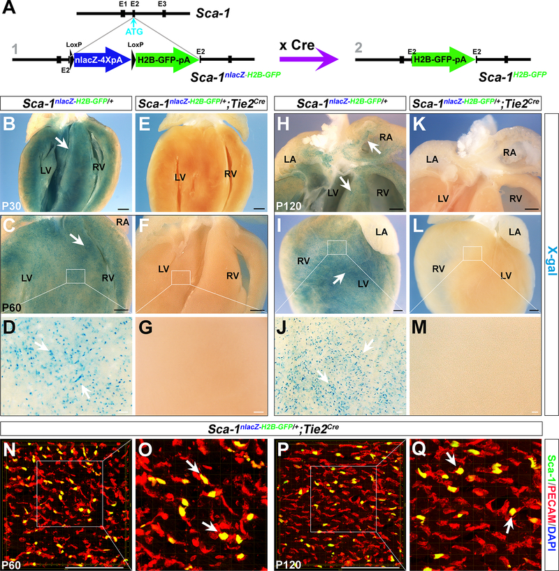
Background: For more than a decade, Sca-1+ cells within the mouse heart have been widely recognized as a stem cell population with multipotency that can give rise to cardiomyocytes, endothelial cells, and smooth muscle cells in vitro and after cardiac grafting. However, the developmental origin and authentic nature of these cells remain elusive.

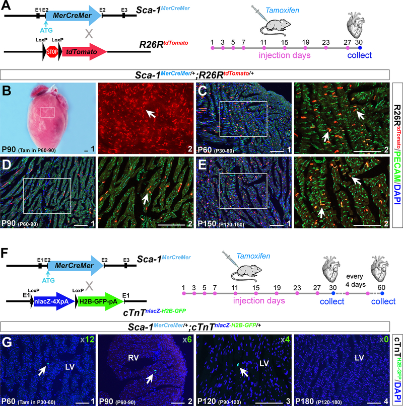
Methods: Here, we used a series of high-fidelity genetic mouse models to characterize the identity and regenerative potential of cardiac resident Sca-1+ cells.

Results: With these novel genetic tools, we found that Sca-1 does not label cardiac precursor cells during early embryonic heart formation. Postnatal cardiac resident Sca-1+ cells are in fact a pure endothelial cell population. They retain endothelial properties and exhibit minimal cardiomyogenic potential during development, normal aging and upon ischemic injury.

Conclusions: Our study provides definitive insights into the nature of cardiac resident Sca-1+ cells. The observations challenge the current dogma that cardiac resident Sca-1+ cells are intrinsic stem cells for myocardial development, renewal, and repair, and suggest that the mechanisms of transplanted Sca-1+ cells in heart repair need to be reassessed.

Keywords: heart failure; regeneration; stem cells.


Conflict of interest statement

Conflicts of Interest Disclosures: None.

Competing financial interests: The authors declare no competing financial interests.

Figures

Figure 1.
Figure 1.. Heterogeneity of cardiac resident Sca-1+ cells.
(A) Diagram of the Sca-1H2B-tdTomato/+;c-KitH2B-GFP/+ double heterozygous alleles. (B-C) Longitudinal sections of Sca-1H2B-tdTomato/+;c-KitH2B-GFP/+ mouse hearts at P30 (B) and P120 (C). Partial Sca-1H2B-tdTomato cells and c-KitH2B-GFP cells showed co-localization (yellow, arrows in B2 and C2). (D) Flow cytometry analysis of 4-month-old Sca-1H2B-tdTomato/+;c-KitH2B-GFP/+ mouse heart cells. (E) Diagram of the Sca-1H2B-tdTomato/+;PDGFRaH2B-GFP/+ double heterozygous alleles. (F-G) Longitudinal sections of Sca-1H2B-tdTomato/+;PDGFRaH2B-GFP/+ mouse hearts at P60 (F) and P240 (G). Some Sca-1H2B-tdTomato cells and PDGFRaH2B-GFP cells showed co-localization (yellow, arrows in F2 and G2). (H) Flow cytometry analysis of 4-month-old Sca-1H2B-tdTomato/+;PDGFRaH2B-GFP/+ mouse heart cells. (I) Diagram of the Sca-1H2B-tdTomato/+;Nkx2.5H2B-GFP/+ double heterozygous alleles. (J, K) Longitudinal sections of Sca-1H2B-tdTomato/+;Nkx2.5H2B-GFP/+ mouse hearts at P30 (M) and P120 (N). Sca-1H2B-tdTomato cells (red, arrows in J2 and K2) and NKx2.5H2B-GFP-positive cells (green, arrowheads in M2 and N2) were not co-localized. (L) Diagram of the Sca-1H2B-tdTomato/+;cTnTH2B-GFP/+ double heterozygous alleles. (M, N) Longitudinal sections of Sca-1H2B-tdTomato/+;cTnTH2B-GFP/+ mouse hearts at P30 (J) and P120 (K). No Sca-1H2B-tdTomato cells (red, arrows in M2 and N2) were cTnTH2B-GFP-positive (green, arrowheads in J2 and K2). LV, left ventricle; RV, right ventricle. n=3 for each stage. Scale bar, 100 μm.
Figure 2.
Figure 2.. Cardiac resident Sca-1+ cells are of the Tie2 lineage.
(A) Diagram of the Sca-1nLacZ-H2B-GFP/+ reporter allele. Sca-1H2B-GFP is expressed when the nlacZ cassette is removed by Cre excision. (B-M) X-gal staining of hearts from Sca-1nLacZ-H2B-GFP/+ and Sca-1nLacZ-H2B-GFP/+;Tie2Cre/+ littermate mice at P30 (B, E), P60 (C, F) and P120 (H-L). D, G, J and M are high-magnification photomicrographs corresponding with the areas outlined in C, F, I and L (white rectangle). Numerous Sca-1nLacZ-positive cells were observed in Sca-1nLacZ-H2B-GFP/+ hearts (arrows in B, C D, H, I and J), but no X-gal-positive cells were seen in Sca-1nLacZ-H2B-GFP/+;Tie2Cre/+ hearts. (N-Q) Z-stack images of immunostaining with an anti-PECAM antibody (red) of Sca-1nLacZ-H2B-GFP/+;Tie2Cre/+ hearts at P60 (N, O) and P120 (P, Q). Sca-1H2B-GFP-positive cells co-localized with PECAM (yellow, arrows in O and Q). LA, left atria; LV, left ventricle; RA, right atria; RV, right ventricle. n=3 for each stage. Black scale bar, 1 mm. White scale bar, 100 μm.
Figure 3.
Figure 3.. Sca-1+ cells in the adult heart have minimal myogenic potential.
(A) Diagram of the Sca-1MerCreMer/+ allele. Sca-1MerCreMer/+ mice were crossed with the ROSA26RtdTomato reporter to obtain Sca-1MerCreMer/+;ROSA26RtdTomato/+ double heterozygous mice. Tamoxifen was administered 9 times in one month (days 1, 3, 5, 7, 11, 15, 19, 23 and 27) to induce Sca-1MerCreMer expression. (B) A representative Sca-1MerCreMer/+;ROSA26RtdTomato/+ heart showed substantial ROSA26RtdTomato-positive cells present after tamoxifen treatment (red, arrows in B2). (C-E) Immunostaining with an anti-PECAM antibody (green) of Sca-1MerCreMer/+;ROSA26RtdTomato/+ hearts at P30, P60 and P90 after 1 month of tamoxifen treatment. ROSA26RtdTomato cells co-localized with PECAM staining in these hearts (yellow, arrows in C2, D2 and E2). (F) Diagram of the Sca-1MerCreMer/+;cTnTnLacZ-H2B-GFP/+ double heterozygous alleles. cTnT2H2B-GFP was expressed when Cre activity was specifically induced in cardiomyocytes. Hearts were collected after 1 month of tamoxifen treatment. (G) All cryosections (10 μm) of Sca-1MerCreMer/+;cTnTnLacZ-H2B-GFP/+ mouse hearts at P60, P90, P120 and P180 were examined under a microscope. Very few GFP cells were detected (arrows in G), and the total number was determined and is shown in the corner. LV, left ventricle; RV, right ventricle. Black scale bar, 1 mm. White scale bar, 100 μm.
Figure 4.
Figure 4.. Resident Sca-1+ cells do not convert into cardiomyocytes upon injury.
(A) Longitudinal sections of Sca-1H2B-tdTomato/+ mouse hearts showing that Sca-1+ cells (arrow) are present in the injured area at 5 days post-surgery (dps). Sections were counterstained with DAPI. The infarcted region is indicated by asterisks. (B-D) Sca-1H2B-tdTomato/+;Nkx2.5H2B-GFP/+ mouse hearts were collected at 2 dps (B), 5 dps (C) and 8 dps (D). No Sca-1H2B-tdTomato (arrows in B, C and D) and Nkx2.5H2B-GFP (arrowheads in B, C and D) double-positive cells were found in the infarcted area. (E-G) Sca-1H2B-tdTomato/+;cTnTH2B-GFP/+ mouse hearts were collected at 5 dps (E), 8 dps (F) and 15 dps (G). Sca-1H2B-tdTomato cells in the injured area (arrows in E, F and G) were not co-localized with cTnTH2B-GFP cells (arrowheads in E, F and G). (H) Diagram of time points for LAD surgery and tamoxifen administration for Sca-1MereCreMer/+;cTnTnlacZ-H2B-GFP/+;ROSA26RtdTomato/+ triple heterozygous mice. (I-J) Representative whole-mount view of Sca-1MereCreMer/+;cTnTnlacZ-H2B-GFP/+;ROSA26RtdTomato/+ mouse heart at 60 dps (I). ROSA26RtdTomato-positive cells were detected throughout the heart, including the infarcted region (arrows in J3). J2 and J3 are high-magnification images of the border zone (J2) and infarcted area (J3) in J1. Asterisks in J1–3 indicate the infarcted region. (K-M) Longitudinal sections of Sca-1MereCreMer/+;cTnTnlacZ-H2B-GFP/+;ROSA26RtdTomato/+ hearts at 30 dps (K), 60 dps (L) and 120 dps (M). ROSA26RtdTomato cells were detected in the infarcted region (arrows in K-M). No cTnTH2B-GFP-positive cells were detected in the infarcted region at 30, 60 and 120 dps. M2 is the green fluorescence filter image of M1 and shows the absence of cTnTH2B-GFP-positive cells. (N-O) Sca-1H2B-GFP-positive endothelial cells in the infarcted region of Sca-1nLacZ-H2B-GFP/+;Tie2Cre hearts at 30 dps. N2 and O2 are high-magnification photomicrographs corresponding to the areas outlined in N1 and O1. Black scale bar, 1 mm. White scale bar, 100 μm.

Comment in

References

    1. Passier R, van Laake LW and Mummery CL. Stem-cell-based therapy and lessons from the heart. Nature. 2008;453:322–329. - PubMed
    1. Segers VF and Lee RT. Stem-cell therapy for cardiac disease. Nature. 2008;451:937–942. - PubMed
    1. Laflamme MA and Murry CE. Heart regeneration. Nature. 2011;473:326–335. - PMC - PubMed
    1. Ptaszek LM, Mansour M, Ruskin JN and Chien KR. Towards regenerative therapy for cardiac disease. Lancet. 2012;379:933–942. - PubMed
    1. van Berlo JH and Molkentin JD. An emerging consensus on cardiac regeneration. Nat Med. 2014;20:1386–93. - PMC - PubMed

Publication types

MeSH terms