Clinical Trial
. 2019 Feb 15;124(4):539-552.
doi: 10.1161/CIRCRESAHA.118.314050.

Predicting Patient Response to the Antiarrhythmic Mexiletine Based on Genetic Variation

Affiliations
Clinical Trial

Predicting Patient Response to the Antiarrhythmic Mexiletine Based on Genetic Variation

Wandi Zhu et al. Circ Res. .

Abstract

Rationale: Mutations in the SCN5A gene, encoding the α subunit of the Nav1.5 channel, cause a life-threatening form of cardiac arrhythmia, long QT syndrome type 3 (LQT3). Mexiletine, which is structurally related to the Na+ channel-blocking anesthetic lidocaine, is used to treat LQT3 patients. However, the patient response is variable, depending on the genetic mutation in SCN5A.

Objective: The goal of this study is to understand the molecular basis of patients' variable responses and build a predictive statistical model that can be used to personalize mexiletine treatment based on patient's genetic variant.

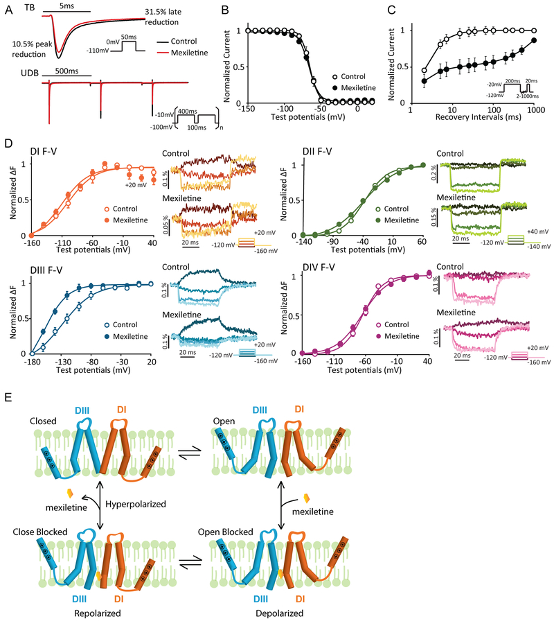
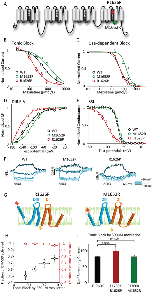
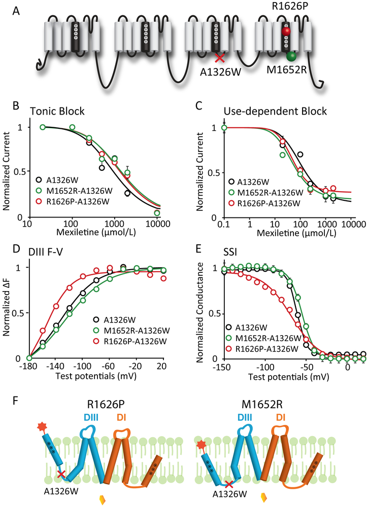
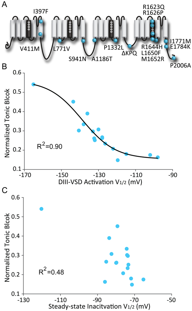
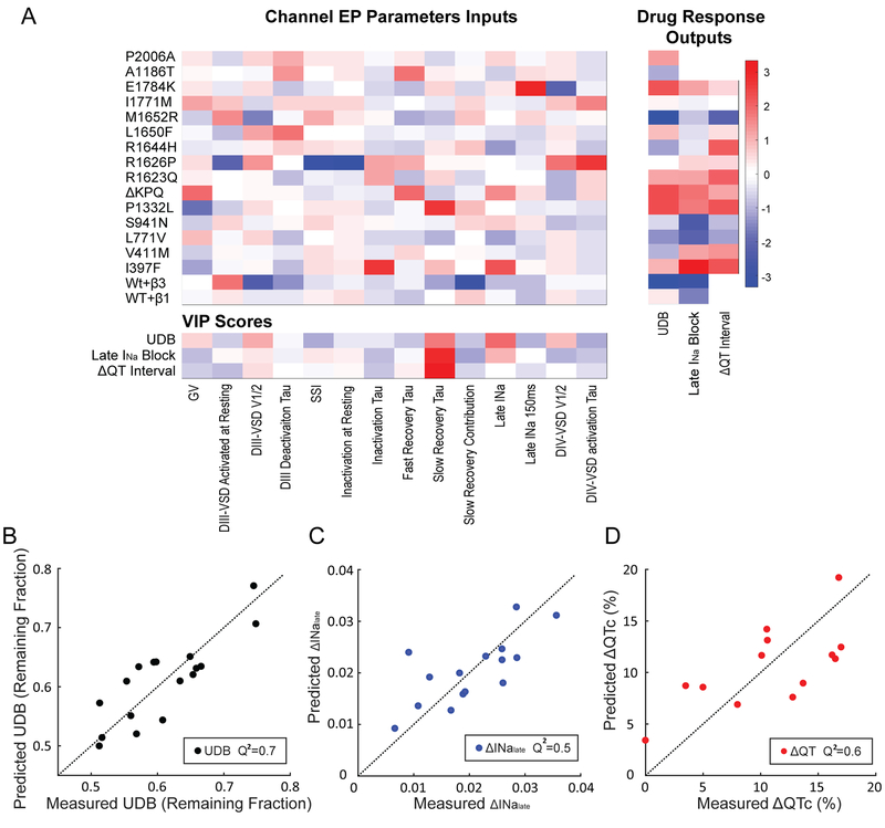
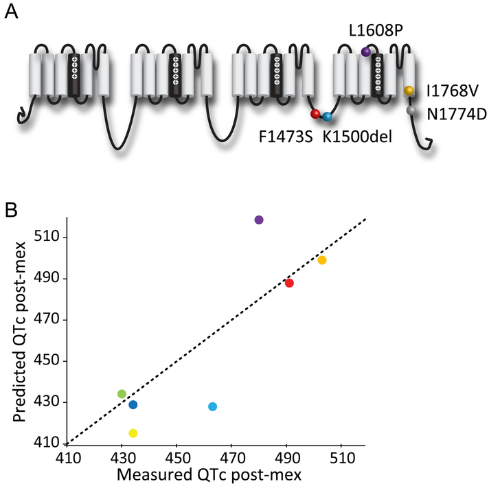
Methods and results: We monitored the cardiac Na+ channel voltage-sensing domain (VSD) conformational dynamics simultaneously with other gating properties for the LQT3 variants. To systematically identify the relationship between mexiletine block and channel biophysical properties, we used a system-based statistical modeling approach to connect the multivariate properties to patient phenotype. We found that mexiletine altered the conformation of the Domain III VSD, which is the same VSD that many tested LQT3 mutations affect. Analysis of 15 LQT3 variants showed a strong correlation between the activation of the Domain III-VSD and the strength of the inhibition of the channel by mexiletine. Based on this improved molecular-level understanding, we generated a systems-based model based on a dataset of 32 LQT3 patients, which then successfully predicted the response of 7 out of 8 patients to mexiletine in a blinded, retrospective trial.

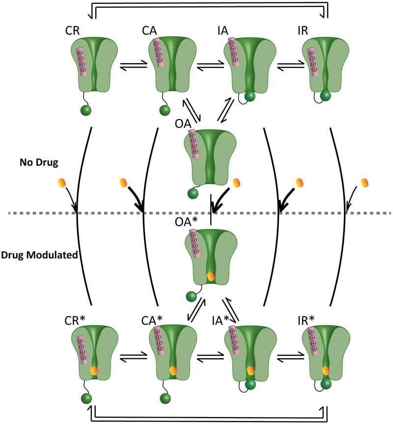
Conclusions: Our results imply that the modulated receptor theory of local anesthetic action, which confines local anesthetic binding effects to the channel pore, should be revised to include drug interaction with the Domain III-VSD. Using an algorithm that incorporates this mode of action, we can predict patient-specific responses to mexiletine, improving therapeutic decision making.

Keywords: electrophysiology; ion channels; long QT syndrome; mexiletine; precision medicine.


Figures

Figure 1:
Figure 1:. Mexiletine blockade of NaV1.5 channel stabilize the DIII-VSD at the activated position.
A. Representative current traces before and after 250 μM mexiletine tonic block (TB) and use-dependent block (UDB). Comparison between traces before and after mexiletine shows that mexiletine reduces the peak current by 10.5%, but the later component (10ms after peak) by 31.5%. 250 μM mexiletine was used, because channels exhibit moderate TB and UDB at this concentration. B. Steady-state inactivation (SSI) curves before and after 2 mM mexiletine (n=4). Channel SSI was tested by holding the cells from −150 to 20 mV with a 10-mV increment for 200 ms. Fraction of channels available were then measured by peak currents induced by a −20mV test pulse. Mexiletine induces minimal hyperpolarizing shift in SSI curve. C. Channel recovery from inactivation curves before and after 250 μM mexiletine (n=3). Cells were first depolarized to −20 mV to induce inactivation, then allowed to recover at −120 mV for various durations. Fraction of channels recovered were then tested with a −20mV pulse. Mexiletine slows down both phases of recovery, especially the slow recovery. D. Left panels: Voltage dependence of steady-state fluorescence (F-V curves) from four domains (DI-V215C, DII-S805C, DIII-M1296C, DIV-S1618C) before and after 4 mM mexiletine. The mean ± SEM is reported for groups of 4 to 8 cells. Fluorescence after mexiletine was measured after 80% tonic block. Right panels: representative fluorescence traces before and after mexiletine. Four voltage steps ranging from −160 to 20mV (DI, DIII, and DIV) or −140 to 40mV (DII) at a 40mV interval are shown. Mexiletine only affects DIII-VSD by causing a hyperpolarizing shift in DIII F-V curve and slows down DIII-VSD deactivation, without affecting other three domains. E. Proposed schematic (adapted from Arcisio-Miranda lidocaine model) showing the mechanism of mexiletine stabilization of activated DIII-VSD. Only DI and DIII are shown and the VSDs are represented by a single S4 segment for clarity.
Figure 2:
Figure 2:. LQT variants with different sensitivities to mexiletine have distinct voltage dependence of DIII-VSD activation.
A. Topology of Nav1.5 channel and location of the two LQT mutations with distinct mexiletine sensitivity, R1626P (red ball, sensitive) and M1652R (green ball, insensitive). B. Concentration dependence of tonic block (TB) by mexiletine for WT, R1626P, and M1652R channels expressed in Xenopus oocytes (n=3 tested for each drug condition). EC50 values were 761 μM for WT, 2035 μM for M1652R, and 211 μM for R1626P channels. C. Concentration dependence of use-dependent block (UDB) by mexiletine (n=3 tested for each drug condition). Currents were normalized to the peak current elicited by the first depolarizing pulse. EC50 values were 58 μM for WT, 193 μM for M1652R, and 57 μM for R1626P channels. D. Voltage dependence of steady-state fluorescence of DIII. The mean ± SEM is reported for groups of 3 to 4 cells. DIII F-V curve of M1652R showed depolarizing shift, while R1626P showed hyperpolarizing shift compared to WT channels. E. Steady-state inactivation (SSI) curves of WT, R1626P, and M1652R channels (n=3 tested for each variant). F. Representative DIII fluorescence traces from WT-M1296C, M1652R-M1296C, and R1626P-M1296C. All three constructs exhibit distinct fluorescence kinetics and voltage-dependence. G. Proposed schematic showing possible mechanisms underlying the difference in mexiletine sensitivities between R1626P and M1652R. The DIII-VSD in the upward position represents the activated conformation. The lower position represents the inactivated conformation. At resting potential, R1626P has more activated DIII-VSD, which is coupled to the DIII pore domain (S5, S6), causing the pore to remain in a conformation with increase accessibility for mexiletine. In contrast, insensitive M1652R fewer activated DIII-VSDs, causing the DIII-pore to enter a conformation with less accessibility. H. The relationships between % of block and the fraction of DIII-VSD activated, or the fraction of current available for four different holding potentials (−120, −110, −100, −90 mV) (n=3 tested for each holding potential). The fraction of current availability for four potentials are not significantly different from each other. The fraction of the DIII-VSD activated shows a linear relationship with the % of TB. I. TB by 500 μM mexiletine for F1760K, R1626P F1760K, M1652R F1760K channels (n=4 tested for each variant). TBs are not significantly different examined with Mann-Whitney U test, suggesting that the F1760K eliminates LQT variant-dependent mexiletine sensitivity.
Figure 3:
Figure 3:. Mutation that decouples the DIII-VSD from DIII-pore eliminates differences in mexiletine sensitivity among LQT3 variants.
A. Locations of the decoupling mutation A1326W and two LQT3 variant mutations, R1626P and M1652R. A1326W resides on the S4-S5 linker of DIII, a motif that is known to regulate energetic coupling between the VSD and pore. B. Concentration dependence of TB for A1326W, M1652R-A1326W, and R1626P-A1326W channels (n=3 for each drug condition). EC50 values were 965 μM for WT, 1562 μM for M1652R, and 1441 μM for R1626P channels. C. Concentration dependence of UDB for M1652R-A1326W, and R1626P-A1326W channels (n=3 tested for each drug condition). EC50 values were 113 μM for WT, 51 μM for M1652R, and 52 μM for R1626P channels. D. Voltage dependence of steady-state fluorescence of DIII for A1326W, M1652R-A1326W, and R1626P-A1326W channels (n=4 tested for each variant). The differences in voltage dependence of DIII-VSD activation is similar with the A1326W as a background mutation. DIII F-V curve of M1652R-A1326W still showed depolarizing shift, while R1626P-A1326W showed hyperpolarizing shift compared to A1326W channels. E. Steady-state inactivation (SSI) curves of WT, R1626P, and M1652R channels (n=4 tested for each variant). The differences in SSI among different mutations are also preserved in presence of the A1326W background mutation. F. Proposed schematic showing a model of how A1326W eliminates the different sensitivities among LQT variants.
Figure 4:
Figure 4:. Voltage dependence of DIII-VSD activation strongly correlates with tonic block by mexiletine.
A. Locations along the primary sequence and channel topology of 15 LQT3 variants tested. B. Relationship between the voltage dependence of DIII-VSD activation (V1/2 of DIII F-V) and normalized tonic block by mexiletine. The mean ± SEM is reported for groups of 3 to 4 cells. The data were fitted with a Boltzmann function and the correlation calculated. A strong correlation (R2=0.9) between these two parameters were observed when fitted with a Boltzmann function. C. Relationship between the SSI (V1/2 of SSI) and normalized tonic block by mexiletine. The mean ± SEM is reported for groups of 3 to 4 cells. The two parameters are not well-correlated, suggesting that channel inactivation is not a good predictor of mexiletine tonic block.
Figure 5:
Figure 5:. Partial least square (PLS) regression model can predict UDB and QTc shortening by mexiletine from channel gating parameters.
A. Left: heatmap of 14 quantified electrophysiological parameters (EP) of the gating for 15 LQT3 variants and WT channels with β1 or β3 subunits. Right: heatmap of each channel’s responses to mexiletine, including UDB, late INa block and QTc shortening (ΔQTc) in LQT3 patients undergoing mexiletine treatment. EP parameters and mexiletine blockade were reported as the mean for groups of 3-4 cells. Bottom: VIP scores for each gating parameter. VIP scores were ranked by each parameter’s impact on model fitness. Each gating parameter is removed individually, and PLSR model was constructed with the rest of parameters. The corresponding model fitness was calculated based on Q2 of measured block and predicted block with leave-one-out cross-validation. Higher VIP score (red) suggests that the gating parameter is more important for improving the model fitness. B-D. Relationship between measured and predicted UDB, late INa block or ΔQTc. The predictions were made using the PLS regression model with selected parameters with high VIP scores. Model stability was tested with leave-one-out cross validation.
Figure 6:
Figure 6:. PLS regression model predicts QTc shortening by mexiletine for genetic variants
A. Locations of 5 LQT3 variants that are included in the clinical trial and were not used for training the model. B. Comparison of the measured patients’ QTc after mexiletine therapy and the predicted QTc after mexiletine using the PLS regression model.
Figure 7:
Figure 7:. Proposed updated VSD-modulated receptor model for Class Ib antiarrhythmics.
The model comprises five states, CR, CA, OA, IA, and IR. R and A represent the DIII-VSD at resting and activated positions, respectively. C, O, and I represent closed, open and inactivated states of the pore. Mexiletine has different binding affinity to each state. Thick arrows represent high binding affinity, and the thin black arrows represent low binding affinity. After mexiletine blocks the pore, channels enter five drug modulated states, CR*, CA*, OA*, IA*, and IR*. The transition rates between modulated states change compared to those between the unmodulated states. This model provides a molecular basis for how mexiletine preferentially blocks the channel when the DIII-VSD is activated (CA, OA, IA).

Comment in

References

    1. Priori SG, Blomström-Lundqvist C, Mazzanti A. 2015 ESC Guidelines for the management of patients with ventricular arrhythmias and the prevention of sudden cardiac death. Eur Heart J [Internet]. 2015;8:746–837. Available from: http://www.ncbi.nlm.nih.gov/pubmed/16935866
    1. Mazzanti A, Maragna R, Faragli A, Monteforte N, Bloise R, Memmi M, Novelli V, Baiardi P, Bagnardi V, Etheridge SP, Napolitano C, Priori SG. Gene-specific therapy with mexiletine reduces arrhythmic events in patients with long QT syndrome type 3. J Am Coll Cardiol. 2016;67:1053–1058. - PMC - PubMed
    1. Zipes DP, Jalife J. Cardiac Electrophysiology: From Cell to Bedside: Sixth Edition. 2013.
    1. Ruan Y, Liu N, Bloise R, Napolitano C, Priori SG. Gating properties of SCN5A mutations and the response to mexiletine in long-QT syndrome type 3 patients. Circulation. 2007;116:1137–1144. - PubMed
    1. Echt DS, Liebson PR, Mitchell LB, Peters RW, Obias-Manno D, Barker AH, Arensberg D, Baker A, Friedman L, Greene HL, Huther ML, Richardson DW. Mortality and Morbidity in Patients Receiving Encainide, Flecainide, or Placebo. N Engl J Med [Internet]. 1991;324:781–788. Available from: http://www.nejm.org/doi/abs/10.1056/NEJM199103213241201 - DOI - PubMed

Publication types

MeSH terms