. 2018 Dec 19;14(12):e1007623.
doi: 10.1371/journal.pgen.1007623. eCollection 2018 Dec.

AMPK signaling linked to the schizophrenia-associated 1q21.1 deletion is required for neuronal and sleep maintenance

Affiliations

AMPK signaling linked to the schizophrenia-associated 1q21.1 deletion is required for neuronal and sleep maintenance

Stanislav Nagy et al. PLoS Genet. .

Abstract

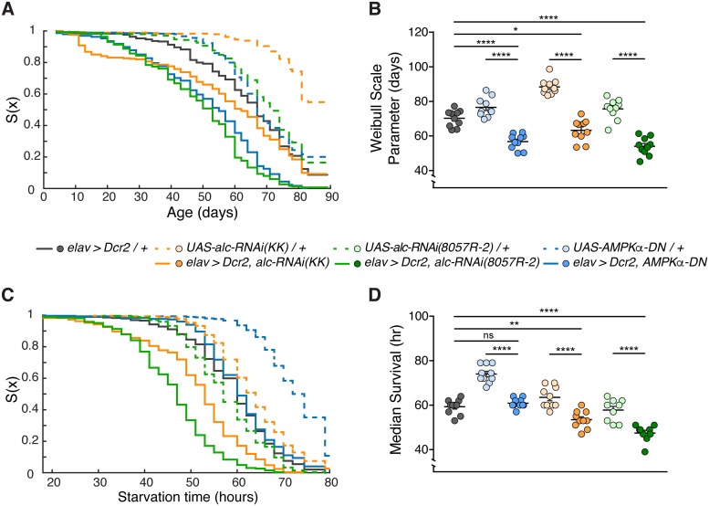
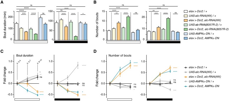
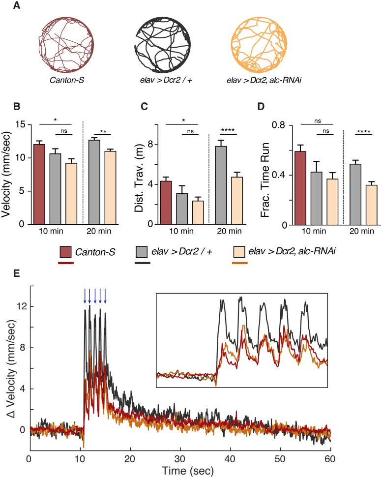
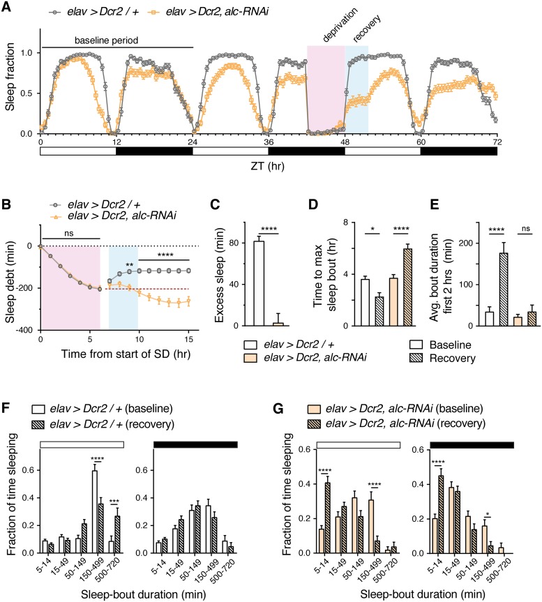
The human 1q21.1 deletion of ten genes is associated with increased risk of schizophrenia. This deletion involves the β-subunit of the AMP-activated protein kinase (AMPK) complex, a key energy sensor in the cell. Although neurons have a high demand for energy and low capacity to store nutrients, the role of AMPK in neuronal physiology is poorly defined. Here we show that AMPK is important in the nervous system for maintaining neuronal integrity and for stress survival and longevity in Drosophila. To understand the impact of this signaling system on behavior and its potential contribution to the 1q21.1 deletion syndrome, we focused on sleep, an important role of which is proposed to be the reestablishment of neuronal energy levels that are diminished during energy-demanding wakefulness. Sleep disturbances are one of the most common problems affecting individuals with psychiatric disorders. We show that AMPK is required for maintenance of proper sleep architecture and for sleep recovery following sleep deprivation. Neuronal AMPKβ loss specifically leads to sleep fragmentation and causes dysregulation of genes believed to play a role in sleep homeostasis. Our data also suggest that AMPKβ loss may contribute to the increased risk of developing mental disorders and sleep disturbances associated with the human 1q21.1 deletion.


Conflict of interest statement

The authors have declared that no competing interests exist.

Figures

Fig 1
Fig 1. Loss of AMPK in the nervous system reduces overall lifespan.
(A) Survival graph showing overall survival for an 89-day period, for male flies raised and kept on standard food. Results obtained from 10 replicates per genotype, each containing 30 flies. (B) Quantification of overall survival. Knockdown of alc or overexpression of AMPKα-DN in the nervous system reduces overall longevity. Survival analyzed using Kaplan-Meier nonparametric method. Survival quantified by fitting each replicate by a Weibull distribution function, right-censoring animals alive at end of experiment. (C) Survival graph showing overall survival for an 80-hour period, for 3-5-day-old male flies raised on normal food and starved on 2% agar alone. Results were obtained from 9–10 replicates per genotype, each containing 30 flies (n > 270). (D) Quantification of survival following starvation. Pan-neuronal knockdown of alc increases sensitivity to starvation. For controls, lines were crossed to w1118. Error bars indicate SEM. Kruskal-Wallis test with Dunn’s post-hoc testing was used to determine statistical significance: *p<0.05, **p<0.01, ***p<0.001, ****p<0.0001, versus the control.
Fig 2
Fig 2. alc/AMPKβ is required for maintenance of dendritic branching in Class-IV sensory neurons.
(A) Representative images of Class-IV pickpocket (ppk)>GFP-expressing neurons from abdominal segment A2 in control (top) and alc-RNAi knockdown third-instar larvae. Individual neurons are outlined. Arrowheads point to beaded morphology of dendrites characteristic of reduced alc. (B) Quantification of total dendrite length (mm) in both feeding (n = 10) and wandering (n = 3) third-instar larvae shows a significant decrease in total dendrite length when alc is knocked down in class-IV neurons. (C-E) Quantification shows no significant change in neuronal area (mm2) (D); however, knockdown of alc decreases both dendritic branching order (C) and branch number (E). Quantification performed using the TREES Matlab toolbox. For controls, lines were crossed to w1118. Error bars indicate SEM. Unpaired T-test was used to determine statistical significance: *p<0.05, **p<0.01, ***p<0.001, ****p<0.0001, versus the control.
Fig 3
Fig 3. Knockdown of alc/AMPKβ in the nervous system impairs courtship-conditioning learning.
(A) A courtship-conditioning assay was used to determine whether alc is required for courtship learning. Courtship index (percentage of time spent courting) decreased after training (with a mated female) in control animals (77% decrease for elav>Dcr-2/+ and 67% decrease for UAS-alc-RNAi/+), indicating learning in both genotypes. There was no significant difference between the performance of naïve and trained animals lacking alc pan-neuronally. Mann Whitney test was used to determine statistical significance: **p<0.01, ***p<0.001, ****p<0.0001, versus the control. Results were obtained from 11 experimental repeats for each genotype, using 3 animals trials each (n = 33 animals). (B) Calculated learning index for alc knockdown and control animals. Error bars indicate SEM. Significance was determined using a one-way ANOVA with Tukey’s post-hoc comparison: **p<0.01 versus the control. For controls, lines were crossed to w1118.
Fig 4
Fig 4. Knockdown of alc/AMPKβ in the nervous system disrupts sleep.
(A-B) Activity (A) and sleep (B) profiles over a 24-hour period for control genotypes elav>Dcr-2 (n = 128) and UAS-alc-RNAi/+ (n = 126) versus elav>Dcr-2, alc-RNAi (n = 126). All data obtained from second 24-hour cycle to allow for acclimatization. Activity and sleep are shown in bins of 30 minutes. White and black bars represent ZT time, 12 hours light and 12 hours dark, respectively. (C) Total sleep (min) in flies with pan-neuronal alc knockdown compared to controls. Total sleep is significantly reduced when alc is knocked down in the nervous system. (D) Comparison of activity outside sleep periods between alc knockdown animals and controls shows that pan-neuronal knockdown of alc increases activity during daytime, but not nighttime. (E) Motion-bout length (min) is significantly increased during the day in alc knockdown flies compared to controls. (F) Average sleep-bout length (min) is significantly reduced during both day- and night-time in alc knockdown flies. (G) The number of sleep bouts per day and night increases when alc is knocked down in the nervous system. (H) Duration of the longest sleep bout (min) is significantly shorter in alc knockdown animals than in controls. (I, J) Distribution of length of sleep bouts for control genotypes elav>Dcr-2/+ and UAS-alc-RNAi/+ versus elav>Dcr-2, alc-RNAi during the day (I) and during the night (J). Flies in which alc has been knocked down pan-neuronally only generate a small proportion of sleep bouts of >150 min, whereas the majority of sleep bouts in controls are of >150 min. For controls, lines were crossed to w1118. Error bars indicate SEM. Kruskal-Wallis test with Dunn’s post-hoc testing was used to determine statistical significance: ***p<0.001, ****p<0.0001, versus the control.
Fig 5
Fig 5. Neuronal loss of AMPK activity causes progressive sleep fragmentation.
(A-B) Quantification of average bout durations (A) and average bout numbers (B) during day and night for elav>Dcr-2/+ (n = 32), UAS-alc-RNAi(KK)/+ (n = 32), elav>Dcr-2, alc-RNAi(KK) (n = 31), UAS-alc-RNAi(8057-R2)/+ (n = 32), elav>Dcr-2, alc-RNAi(8057-R2) (n = 32), UAS-AMPKα-DN/+ (n = 32), elav>Dcr-2, AMPKα-DN (n = 31). Sleep was monitored over 10 days, and data from days 2 to 4 was used. Bout durations are significantly reduced and bout numbers significantly increased when alc is knocked down in the nervous system compared to control genotypes, for both RNAi lines. There is no initial difference in either parameter when dominant-negative AMPKα (AMPKα-DN) is overexpressed in the nervous system. (C-D) Graphical representation of the fold change in bout duration (C) and bout number (D) over time (3-day binned) during both day and night, indicated by white and black bars respectively. Shown are data for elav>Dcr-2, alc-RNAi(KK) and elav>Dcr-2, AMPKα-DN compared to control genotypes. Bout duration significantly decreases over time in both alc knockdown and AMPKα-DN overexpression flies. Bout number significantly increases over time for both genotypes. Significance between days 8–10 and days 2–4 is shown. For controls, lines were crossed to w1118. Error bars indicate SEM. Significance was determined using a Kruskal-Wallis test with Dunn’s post-hoc testing: *p<0.05, **p<0.01, ***p<0.001, ****p<0.0001, versus the control.
Fig 6
Fig 6. Knockdown of alc/AMPKβ in the nervous system does not result in hyperactivity or hypersensitivity.
(A) Example tracks of individual animals over a 10-minute recording period. (B-D) Graphs showing average running velocity (>2mm/sec) (B), total distance traveled (C), and fraction of time moving (velocity > 2 mm/sec) (D) for Canton-S, elav>Dcr-2/+, and elav>Dcr-2, alc-RNAi animals for a 10 minute (n = 10) and 20 minute recording period (n = 30). Error bars indicate SEM. Mann Whitney or Kruskal-Wallis test with Dunn’s post-hoc testing was used to determine statistical significance: ***p<0.001, ****p<0.0001, versus the control. (E) Average baseline subtracted velocity for Canton-S, elav>Dcr-2/+, and elav>Dcr-2, alc-RNAi animals centered on delivery of a five pulse mechanical stimulus (blue arrows). Inset box shows a magnified view of the response. N = 80 animals for each genotype and data averaged over five pulse trains separated by 60 seconds.
Fig 7
Fig 7. Alc/AMPKβ is required in the nervous system for recovery sleep following sleep deprivation.
(A) Sleep profile for control genotype elav>Dcr2/+ (n = 54) versus elav>Dcr2, alc-RNAi (n = 52) for a 72-hour period, showing sleep profiles for baseline, sleep deprivation, and recovery days. White and black bars represent ZT time, 12 hours light and 12 hours dark, respectively. A 6-hour sleep-deprivation period is illustrated using pink shading, typical recovery period illustrated with blue shading. (B) Quantification of sleep debt (min) showed that control flies recovered lost sleep in the first 3 hours following sleep deprivation. Significance determined by 2-way ANOVA with Sidak’s multiple comparisons test between control and alc-RNAi animals shown. Dashed line shows maximum sleep debt. (C) Control animals showed a significant excess of sleep (80 minutes) compared to the same time period of the baseline day while alc-RNAi animals did not. Pan-neuronal knock down of alc completely eliminates recovery sleep. (D) Time to maximum sleep bout (hours) is significantly reduced in control flies following sleep deprivation, whereas it is significantly increased in flies lacking alc in the nervous system. (E) Average bout duration in the first two hours of light phase was significantly increased during recovery in control flies, but not in alc knockdown animals. (F-G) Distribution of length of sleep bouts for control genotype elav>Dcr2/+ (F) versus elav>Dcr2, alc-RNAi (G) during the day and during the night, for baseline and recovery days. Unlike control flies, which showed an increase in long sleep bouts (>150 minutes) during the day, alc knockdown animals showed a decrease in long sleep bouts during both the day and the night, following sleep deprivation. For controls, lines were crossed to w1118. Error bars indicate SEM. For C-E, Mann Whitney test was used to determine statistical significance. For F and G, a 2-way ANOVA with Tukey’s post-hoc comparison was used: *p<0.05, **p<0.01, ***p<0.001, ****p<0.0001, versus the control.

References

    1. Fejgin K, Nielsen J, Birknow MR, Bastlund JF, Nielsen V, Lauridsen JB, et al. A mouse model that recapitulates cardinal features of the 15q13.3 microdeletion syndrome including schizophrenia- and epilepsy-related alterations. Biol Psychiatry. 2014;76(2):128–37. 10.1016/j.biopsych.2013.08.014 . - DOI - PubMed
    1. Malhotra D, Sebat J. CNVs: harbingers of a rare variant revolution in psychiatric genetics. Cell. 2012;148(6):1223–41. 10.1016/j.cell.2012.02.039 . - DOI - PMC - PubMed
    1. Ingason A, Giegling I, Cichon S, Hansen T, Rasmussen HB, Nielsen J, et al. A large replication study and meta-analysis in European samples provides further support for association of AHI1 markers with schizophrenia. Hum Mol Genet. 2010;19(7):1379–86. Epub 2010/01/15. 10.1093/hmg/ddq009 . - DOI - PMC - PubMed
    1. Stefansson H, Rujescu D, Cichon S, Pietilainen OP, Ingason A, Steinberg S, et al. Large recurrent microdeletions associated with schizophrenia. Nature. 2008;455(7210):232–6. Epub 2008/08/01. 10.1038/nature07229 . - DOI - PMC - PubMed
    1. Szelenberger W, Soldatos C. Sleep disorders in psychiatric practice. World Psychiatry. 2005;4(3):186–90. . - PMC - PubMed

Publication types

MeSH terms

Substances

Supplementary concepts

LinkOut - more resources