. 2018 Dec 18;25(12):3530-3542.e5.
doi: 10.1016/j.celrep.2018.11.086.

A Cellular Anatomy of the Normal Adult Human Prostate and Prostatic Urethra

Affiliations

A Cellular Anatomy of the Normal Adult Human Prostate and Prostatic Urethra

Gervaise H Henry et al. Cell Rep. .

Abstract

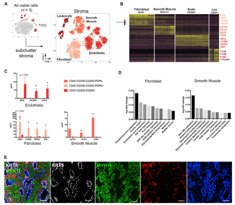
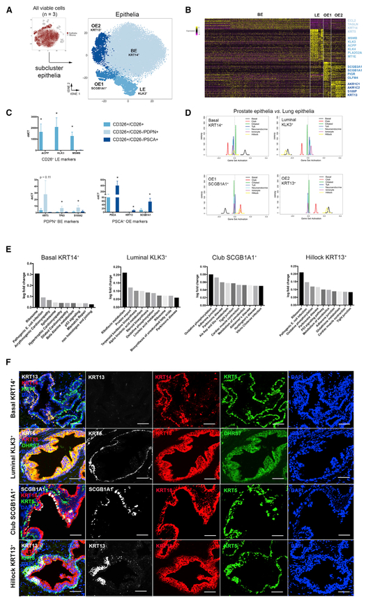
A comprehensive cellular anatomy of normal human prostate is essential for solving the cellular origins of benign prostatic hyperplasia and prostate cancer. The tools used to analyze the contribution of individual cell types are not robust. We provide a cellular atlas of the young adult human prostate and prostatic urethra using an iterative process of single-cell RNA sequencing (scRNA-seq) and flow cytometry on ∼98,000 cells taken from different anatomical regions. Immunohistochemistry with newly derived cell type-specific markers revealed the distribution of each epithelial and stromal cell type on whole mounts, revising our understanding of zonal anatomy. Based on discovered cell surface markers, flow cytometry antibody panels were designed to improve the purification of each cell type, with each gate confirmed by scRNA-seq. The molecular classification, anatomical distribution, and purification tools for each cell type in the human prostate create a powerful resource for experimental design in human prostate disease.

Keywords: GUDMAP; benign prostatic hyperplasia; flow cytometry; human cell atlas; human prostate; prostate cancer; prostate epithelia; prostate stroma; single-cell RNA sequencing; zonal anatomy.


Conflict of interest statement

DECLARATION OF INTERESTS

The authors declare no competing interests.

Figures

Figure 1.
Figure 1.. Identification of Human Prostate Cell Clusters with Bulk and Single-Cell RNA Sequencing
(A) Schematic of human tissue collection and processing for bulk and single-cell RNA sequencing. (B) Aggregated single-cell RNA sequencing (scRNA-seq) data from three organ donor prostate specimens with subclustering into stroma, epithelia, and unknown lineages based on correlation with bulk sequencing data (Figure S2). Clusters were identified and re-merged. (C) Dot plot of cluster-specific genes after in silico removal of stressed cells and supervised identification of neuroendocrine epithelia.
Figure 2.
Figure 2.. Optimization of Flow Cytometry for Purification of Stromal and Epithelial Subtypes
(A) Standard flow cytometry strategy for purification of prostate stroma and epithelial subtypes. (B) Barcoding of cells from traditional FACS gates shows breakdown of cell types within each gate. (C) Quantification of cells within barcoded FACS gates. (D) (Left) CD200 labels 93% of endothelia that CD31 labels. (Right) Podoplanin (PDPN) and CD200 separate endothelia (CD200+), fibroblasts (PDPN+), and smooth muscle (PDPN). (E) PSCA was identified as a potential cell surface marker capable of isolating other epithelial cells after CD26+ luminal epithelia are removed. (F) scRNA-seq of modified FACS gates on a new organ donor prostate specimen is used to demonstrate the increased purity of isolated stromal and epithelial cell types compared to traditional gates in (A)–(C). These data are quantitated in (G).
Figure 3.
Figure 3.. Identification and Isolation of Pure Stromal Subtypes in the Normal Human Prostate
(A) scRNA-seq data aggregated from three normal prostate specimens subclustered into the stromal lineage. (B) Heatmap of the top 100 differentially expressed genes in each stromal subcluster with highlighted DEGs, suggesting putative identities. (C) qPCR of FACS-isolated stromal subtypes from three organ donor prostate specimens demonstrates the enrichment of cell type-specific DEGs. (D) Gene set enrichment analysis (GSEA) of non-endothelial stromal populations compared to KEGG pathways. (E) Immunofluorescent labeling of smooth muscle (MYH11), fibroblasts (DCN), and basal epithelia (KRT5). *p ≤ 0.05; Scale bar, 100 μm. qPCR data are represented as mean ± SEM. Statistical significance of qPCR data was calculated by t test.
Figure 4.
Figure 4.. Identification and Isolation of Pure Epithelial Subtypes in the Normal Human Prostate
(A) scRNA-seq data aggregated from three normal human prostate specimens subclustered into the epithelial lineage. (B) Heatmap of the top 100 differentially expressed genes in each epithelial subcluster with highlighted DEGs. (C) qPCR of FACS-isolated epithelial subtypes from three organ donor prostate specimens demonstrates the enrichment of cell type-specific DEGs. (D) GSEA of four human prostate epithelial cell types compared to mouse lung epithelial cell types. (E) KEGG pathways enriched in epithelial cell types. (F) Immunofluorescent labeling of basal epithelia (KRT5), luminal epithelia (DHRS7), club epithelia (SCGB1A1), and hillock epithelia (KRT13). *p ≤ 0.05; Scale bar, 50 μm. qPCR data are represented as mean ± SEM. Statistical significance of qPCR data was calculated by t test.
Figure 5.
Figure 5.. Anatomical Location of Epithelial and Stromal Cell Types in the Normal Human Prostate
(A) Transition and central zones of the prostate were dissected away from the peripheral zone from three young organ donors for scRNA-seq (pre-dissected tissue inset). (B) Quantification of scRNA-seq-identified cell types after segregation by anatomical zone from 3 patients’ aggregated data. (C) Representative FACS analysis of epithelia from transition and peripheral zone tissue from five young organ donors after segregation by anatomical zone. (D) Quantification of FACS data on zonal enrichment of cell types. (E) Immunofluorescence of prostate whole-mount sections displays enrichment of club and hillock epithelial cell types in the central and transition zones and the urethra and a concentration of fibroblasts in the peri-urethral and central zone regions. *p ≤ 0.05; Scale bar, 100 μm. Statistical significance of scRNA-seq (A) and flow cytometry (B) data was calculated by t test.

References

    1. Abler LL, Keil KP, Mehta V, Joshi PS, Schmitz CT, and Vezina CM (2011). A high-resolution molecular atlas of the fetal mouse lower urogenital tract. Dev. Dyn 240, 2364–2377. - PMC - PubMed
    1. Butler A, Hoffman P, Smibert P, Papalexi E, and Satija R (2018). Integrating single-cell transcriptomic data across different conditions, technologies, and species. Nat. Biotechnol 36, 411–420. - PMC - PubMed
    1. Chuang YY, Chen Y, Gadisetti, Chandramouli VR, Cook JA, Coffin D, Tsai MH, DeGraff W, Yan H, Zhao S, et al. (2002). Gene expression after treatment with hydrogen peroxide, menadione, or t-butyl hydroperoxide in breast cancer cells. Cancer Res. 62, 6246–6254. - PubMed
    1. Cunha GR, and Lung B (1978). The possible influence of temporal factors in androgenic responsiveness of urogenital tissue recombinants from wild-type and androgen-insensitive (Tfm) mice. J. Exp. Zool 205, 181–193. - PubMed
    1. Cunha GR, Ricke W, Thomson A, Marker PC, Risbridger G, Hayward SW, Wang YZ, Donjacour AA, and Kurita T (2004). Hormonal, cellular, and molecular regulation of normal and neoplastic prostatic development.J. Steroid Biochem. Mol. Biol 92, 221–236. - PubMed

Publication types