

Resection of ruptured hepatic teratoma in an adult
- PMID: 30567058
- PMCID: PMC6259038
- DOI: 10.1016/j.ijscr.2018.11.032
Resection of ruptured hepatic teratoma in an adult
Abstract
Introduction: Extragonadal locations of teratomas are uncommonly reported in the literature. Teratomas are neoplasms usually found in the gonadal organs: ovaries and testis. The majority of teratomas are found in the pediatric age group. Furthermore, teratomas originating in the liver are exceedingly rare with only 11 case reports in adult populations.
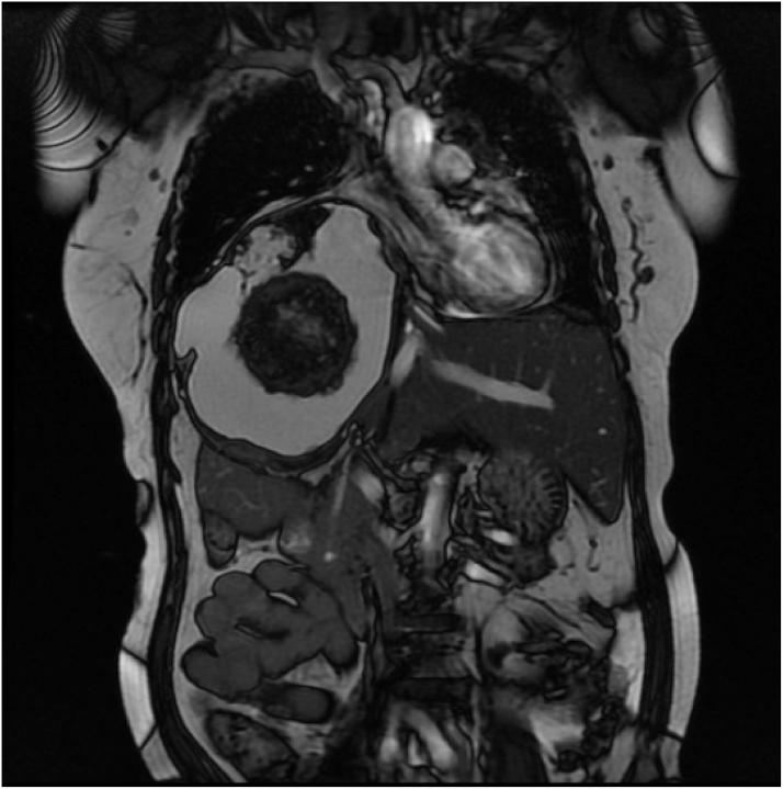
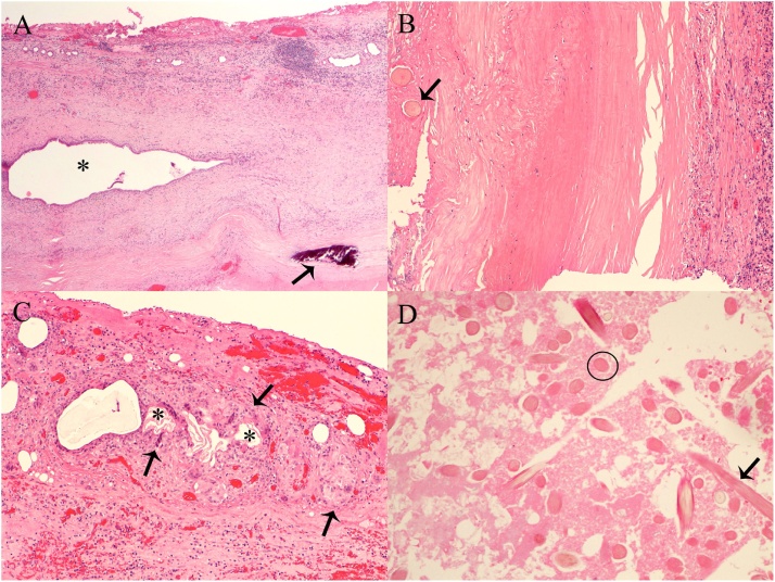
Presentation of case: We present a case of a 65 year-old female who presented to hospital with sudden onset abdominal pain from a centrally located ruptured hepatic teratoma on CT scan. The patient underwent urgent surgery. The diagnosis of cystic mature teratoma was confirmed on histopathology. Patient was discharged on post-operative day 5. At 12 week follow-up, no post-operative complications were identified.
Discussion: Hepatic teratomas are a rarely encountered neoplasm, especially in the adult population. Our case report is unique, as it represents the only clinical presentation of mass rupture in an adult liver teratoma. CT scan identified a well circumscribed mass containing adipose tissue, fluid, and calcifications characteristic of teratoma. Complete surgical resection is mainstay treatment. A definitive diagnosis of a mature teratoma is confirmed by histopathological findings.
Conclusion: Presented is a rare case of ruptured hepatic teratoma in an adult who underwent surgical resection.
Keywords: Case report; General surgery; Hepatic teratoma; Hepatobiliary; Surgery; Teratoma.
Copyright © 2018 The Authors. Published by Elsevier Ltd.. All rights reserved.
Figures


References
-
- Ashley D.J. Origin of teratomas. Cancer. 1973;32(2):390–394. - PubMed
-
- Davidson A.J., Hartman D.S., Goldman S.M. Mature teratoma of the retroperitoneum: radiologic, pathologic, and clinical correlation. Radiology. 1989;172(2):421–425. - PubMed
-
- Martin L.C., Papadatos D., Michaud C., Thomas J. Best cases from the AFIP: liver teratoma. Radiographics. 2004;24:1467–1471. - PubMed
-
- Certo M., Franca M., Gomes M., Machado R. Liver teratoma. Acta Gastroenterol. Belg. 2008;71(2):275–279. - PubMed
LinkOut - more resources
Full Text Sources
Research Materials
