Case Reports
. 2018 Nov 28;11(1):e226611.
doi: 10.1136/bcr-2018-226611.

Eagle's syndrome: a piercing matter

Affiliations
Case Reports

Eagle's syndrome: a piercing matter

Matthew Zammit et al. BMJ Case Rep. .

Abstract

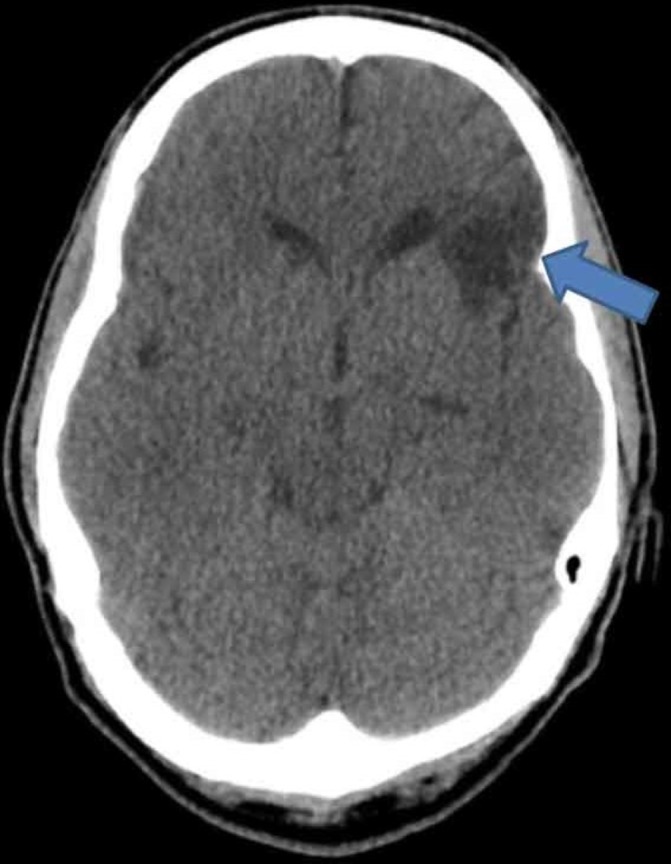
We present an unusual case of Eagle's syndrome with bilateral internal carotid artery dissection in a 45-year-old man. Initial symptomatology included ipsilateral headaches and facial sensory symptoms. A right horner's syndrome was present on clinical examination. Radiological imaging revealed an old infarct, with bilateral carotid dissections and bilateral elongated styloid processes consistent with Eagle's syndrome. Despite initiation of secondary prevention with antiplatelet therapy, he had two further ischaemic events. The case highlights the symptomatology and complications of Eagle's syndrome, with its management discussed through a review of similar case reports.

Keywords: neuroimaging; neurology; stroke.


Conflict of interest statement

Competing interests: None declared.

Figures

Figure 1
Figure 1
CT of the brain displaying an old left frontal lobe infarct, as indicated by the blue arrow.
Figure 2
Figure 2
CT angiography of the aortic arch and neck revealing bilateral internal carotid dissection - shown by the two white arrows.
Figure 3
Figure 3
CT angiography of the aortic arch and neck revealing right internal carotid artery dissection at the level of the blue arrow.
Figure 4
Figure 4
CT angiography of the aortic arch displaying elongated styloid processes, as annotated by the two white arrows.
Figure 5
Figure 5
Diffusion-weighted MRI indicating acute infarction of the right caudate head in the territory of the anterior cerebral artery (white arrow).
Figure 6
Figure 6
Diffusion-weighted MRI revealing right middle cerebral artery territory infarct.

References

    1. Zetterling M, Carlström C, Conrad P. Review article - Internal carotid artery dissection. Acta Neurol Scand 2000;101:1–7. 10.1034/j.1600-0404.2000.00001.x - DOI - PubMed
    1. Mathews T, Achar R, Rattan G. Eagle Syndrome-vascular type-A case presentation and a short review of literature. Case Report 2012. 10.1594/ranzcraocr2012/R-0032 - DOI
    1. Dewan MC, Morone PJ, Zuckerman SL, et al. . Paradoxical ischemia in bilateral Eagle syndrome: a case of false-localization from carotid compression. Clin Neurol Neurosurg 2016;141:30–2. 10.1016/j.clineuro.2015.12.004 - DOI - PubMed
    1. Piagkou M, Anagnostopoulou S, Kouladouros K, et al. . Eagle’s syndrome: a review of the literature. Clin Anat 2009;22:545–58. 10.1002/ca.20804 - DOI - PubMed
    1. Badhey A, Jategaonkar A, Anglin Kovacs AJ, et al. . Eagle syndrome: a comprehensive review. Clin Neurol Neurosurg 2017;159:34–8. 10.1016/j.clineuro.2017.04.021 - DOI - PubMed

Publication types

MeSH terms

Substances

Supplementary concepts