

Rare occurrence of a huge traumatic extradural haematoma in a patient with an ipsilateral sylvian arachnoid cyst
- PMID: 30567181
- PMCID: PMC6301488
- DOI: 10.1136/bcr-2018-227525
Rare occurrence of a huge traumatic extradural haematoma in a patient with an ipsilateral sylvian arachnoid cyst
Abstract
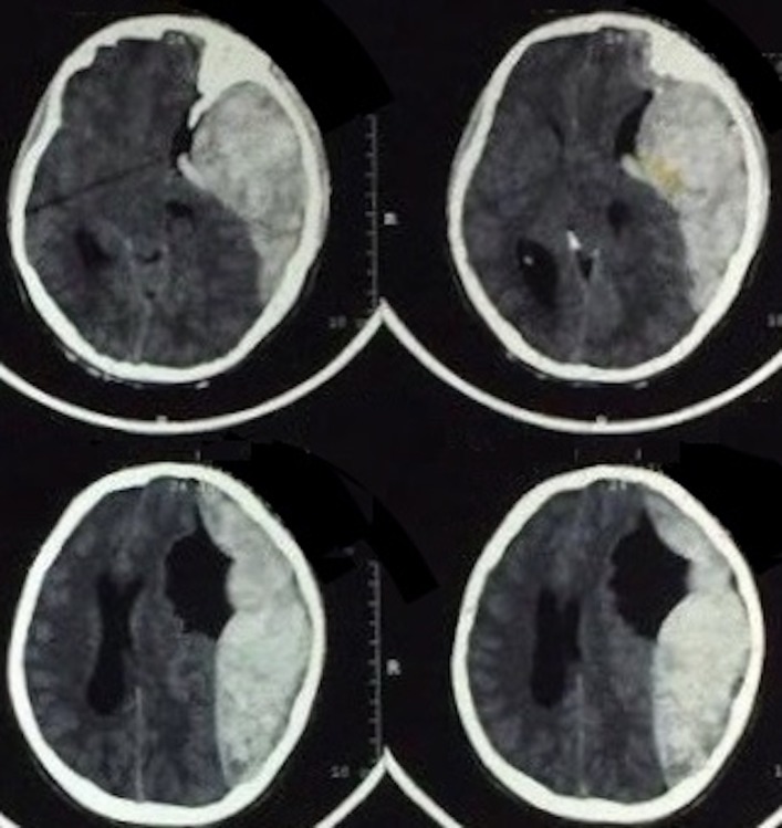
A man, a teenage victim of an assault to the head, presented to the emergency department, in Baghdad, with a Glasgow Coma Score of 4/15 (E1 M2 V1) and total right-sided paralysis. CT of the brain revealed a large-left sided frontotemporoparietal extradural haematoma with the presence of an ipsilateral sylvian arachnoid cyst deep to the haematoma. Urgent surgical evacuation of the haematoma was performed, leaving the arachnoid cyst intact. The patient improved and gained full consciousness within 4 days.Three years after the initial trauma, the patient has remained well. This case required a thorough discussion of the surgical options, in particular whether to intervene with the associated cyst, and whether any intervention with the cyst should be performed in the same or future operations. This dilemma forms the basis of the discussion in the following report.
Keywords: neurosurgery; trauma CNS /PNS.
© BMJ Publishing Group Limited 2018. No commercial re-use. See rights and permissions. Published by BMJ.
Conflict of interest statement
Competing interests: None declared.
Figures


Similar articles
-
Extradural hematoma complicating arachnoid cyst--case report.Zentralbl Neurochir. 1994;55(3):172-4. Zentralbl Neurochir. 1994. PMID: 7810256
-
Arachnoid cyst presenting as an extradural haematoma.Br J Neurosurg. 1991;5(6):635-7. doi: 10.3109/02688699109002888. Br J Neurosurg. 1991. PMID: 1772611
-
Asymptomatic presentation of huge extradural hematoma in a patient with arachnoid cyst.Br J Neurosurg. 2012 Dec;26(6):917-8. doi: 10.3109/02688697.2012.680623. Epub 2012 Aug 21. Br J Neurosurg. 2012. PMID: 22905883
-
Spontaneous resolution of arachnoid cysts: review and features of an unusual case.Acta Neurochir (Wien). 2007 Jan;149(1):75-8; discussion 78. doi: 10.1007/s00701-006-1073-1. Epub 2006 Dec 21. Acta Neurochir (Wien). 2007. PMID: 17180304 Review.
-
[Prepontine epidural hemorrhage].Zentralbl Neurochir. 1998;59(3):185-8. Zentralbl Neurochir. 1998. PMID: 9816670 Review. German.
Cited by
-
PubMed-indexed neurosurgical research productivity of Iraq-based neurosurgeons.Surg Neurol Int. 2021 May 17;12:223. doi: 10.25259/SNI_47_2021. eCollection 2021. Surg Neurol Int. 2021. PMID: 34221554 Free PMC article. Review.
References
-
- Kadioğly HH, Oztürk M, Aydin IH. Extradural hematoma complicating arachnoid cyst–case report. Zentralbl Neurochir 1994;55:172–4. - PubMed
Publication types
MeSH terms
LinkOut - more resources
Full Text Sources