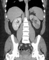
Primitive neuroectodermal tumour with synchronous ipsilateral clear cell carcinoma of the kidney
- PMID: 30567211
- PMCID: PMC6301484
- DOI: 10.1136/bcr-2018-224273
Primitive neuroectodermal tumour with synchronous ipsilateral clear cell carcinoma of the kidney
Abstract
We report the first case of a synchronous ipsilateral primitive neuroectodermal tumour (PNET) and clear cell renal cell carcinoma of the kidney. A 37-year-old man presented to the emergency department with a 24-hour history of colicky abdominal pain and visible haematuria. He had no relevant surgical or medical history. Physical examination was unremarkable apart from mild left flank tenderness. Triphasic CT of the abdomen and pelvis showed two solid lesions in the left kidney. Further staging CT of the chest showed no evidence of local or distal metastasis. He subsequently underwent laparoscopic radical nephrectomy. Pathological analysis of the kidney showed two synchronous renal tumours, a clear cell carcinoma and PNET of the kidney. The patient received adjuvant chemotherapy according to Ewing's sarcoma chemotherapy protocol. Surveillance CT scans at 3, 6 and 12 months showed no evidence of disease recurrence or metastasis.
Keywords: pathology; urological cancer; urological surgery.
© BMJ Publishing Group Limited 2018. No commercial re-use. See rights and permissions. Published by BMJ.
Conflict of interest statement
Competing interests: None declared.
Figures


Similar articles
-
[Primitive neuroectodermal tumor of kidney : a case report].Hinyokika Kiyo. 2013 Jun;59(6):363-7. Hinyokika Kiyo. 2013. PMID: 23827869 Japanese.
-
[Primitive neuroectodermal tumor of the kidney].Urologe A. 2006 Jun;45(6):735-8. doi: 10.1007/s00120-006-1029-3. Urologe A. 2006. PMID: 16534648 German.
-
Ewing's sarcoma / primitive neuroectodermal tumor of the kidney.Aktuelle Urol. 2009 Aug;40(4):247-9. doi: 10.1055/s-0028-1098827. Epub 2009 Mar 17. Aktuelle Urol. 2009. PMID: 19294616
-
[A case of primitive neuroectodermal tumor of the kidney].Nihon Hinyokika Gakkai Zasshi. 1999 Jun;90(6):639-42. doi: 10.5980/jpnjurol1989.90.639. Nihon Hinyokika Gakkai Zasshi. 1999. PMID: 10422440 Review. Japanese.
-
Renal Ewing's sarcoma/primitive neuroectodermal tumor: a case report and literature review.Beijing Da Xue Xue Bao Yi Xue Ban. 2017 Oct 18;49(5):919-923. Beijing Da Xue Xue Bao Yi Xue Ban. 2017. PMID: 29045981 Review.
