Review
. 2018 Dec 18;10(12):723.
doi: 10.3390/v10120723.

Prion Strain-Specific Structure and Pathology: A View from the Perspective of Glycobiology

Affiliations
Review

Prion Strain-Specific Structure and Pathology: A View from the Perspective of Glycobiology

Ilia V Baskakov et al. Viruses. .

Abstract

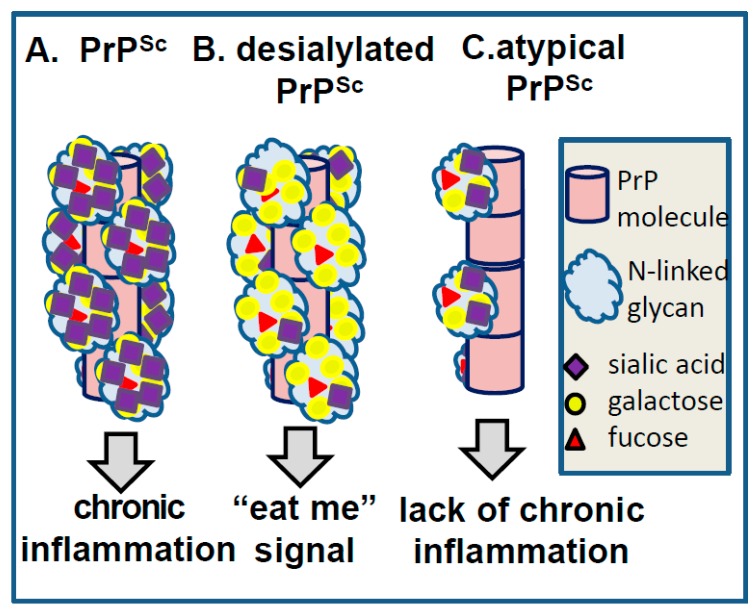
Prion diseases display multiple disease phenotypes characterized by diverse clinical symptoms, different brain regions affected by the disease, distinct cell tropism and diverse PrPSc deposition patterns. The diversity of disease phenotypes within the same host is attributed to the ability of PrPC to acquire multiple, alternative, conformationally distinct, self-replicating PrPSc states referred to as prion strains or subtypes. Structural diversity of PrPSc strains has been well documented, yet the question of how different PrPSc structures elicit multiple disease phenotypes remains poorly understood. The current article reviews emerging evidence suggesting that carbohydrates in the form of sialylated N-linked glycans, which are a constitutive part of PrPSc, are important players in defining strain-specific structures and disease phenotypes. This article introduces a new hypothesis, according to which individual strain-specific PrPSc structures govern selection of PrPC sialoglycoforms that form strain-specific patterns of carbohydrate epitopes on PrPSc surface and contribute to defining the disease phenotype and outcomes.

Keywords: N-linked glycans; glycosylation; prion disease; prion strains; prions; sialic acid; sialylation.


Conflict of interest statement

The authors declare no conflict of interest.

Figures

Figure 1
Figure 1
Staining of PrPSc plaques with SNA lectin. Images of SO5 PrPSc plaques (AC) or atypical PrPSc plaques (E,F) in hamster brains stained with anti-PrP 3F4 antibody (A,E), SNA lectin (B,F), or secondary antibody used for SNA staining as negative control (C). Plaques are shown by arrows. Staining of normal age-matched control with SNA lectin is shown in (D). Brains fixed in 10% neutral buffered formalin were treated with 95% formic acid for 1 h before embedding in paraffin wax and sectioning into 4 µm sections. After a standard rehydration procedure, slides were submerged in 10 mM tri-sodium citrate buffer, pH 6.0, boiled for 5 min by microwaving at 20% power, and cooled for 1 h before proceeding with lectin staining. Incubation in 3% hydrogen peroxide in methanol for 20 min was used to remove endogenous peroxidase activity. After 5 min wash in running water, slides were incubated for 1 h with 5 µg/mL biotin-labeled elderberry bark lectin (SNA, Vector laboratories, Burlingame, CA) diluted in lectin buffer, pH 7.6 (50 mM Tris, 150 mM NaCl, 1 mM MgCl2, 0.75 mM CaCl2). Following triple 5 min wash in lectin buffer, the slides were incubated for 30 min in 5 µg/mL horse radish peroxidase-labeled streptavidin (Thermo Fisher scientific, Waltham, MA), then again washed three times with lectin buffer, and developed with 3,3’ Diaminobenzidine (DAB) Quanto chromogen and substrate (VWR, Radnor, PA).
Figure 2
Figure 2
Schematic diagram illustrating selective recruitment of PrPC sialoglycoforms in a strain-specific manner according to the PrPC sialylation status. The left panel shows distribution of PrPC molecules according to their glycosylation status (in horizontal dimension) and sialylation status (in vertical dimension) ranging from non-sialylated to highly sialylated molecules. PrPC molecules are shown as blue circles and sialic acid residues as red diamonds. The panels on the right show 2D Western blots of three prion strains with different recruitment selectivity. While 263K (strain #1) recruits PrPC sialoglycoforms without strong preferences, hypersialylated PrPC molecules are preferentially excluded from RML (strain #2) and excluded even stronger from atypical PrPSc (strain #3). As a result of strain-specific exclusion of highly sialylated PrPC, ratios of glycoforms within PrPSc shift toward mono- and unglycosylated glycoform, as illustrated by corresponding 1D Western blots. Adapted from Baskakov and Katorcha 2016 [35].
Figure 3
Figure 3
Correlation between strain-specific sialylation status and glycoform ratio. Strain-specific percentages of diglycosylated glycoforms plotted as a function of strain-specific percentage of hypersialylated glycoforms within PrPSc. Mean and standard deviations are shown (n = 3 animals). Black solid line shows the result of linear fitting of the percent of diglycosylated as a function of the percent of hypersialylated for brain-derived PrPSc. Adapted from Katorcha et al. 2015 [37].
Figure 4
Figure 4
Schematic diagram illustrating differences in quaternary assembly between non-selective (left panels) and selective (right panels) strains. It is proposed that non-selective strains can accommodate diglycosylated sialoglycoforms because of rotation between neighboring PrP molecules that allows spatial separation of glycans and reduces electrostatic repulsion. In selective strains, the rotation between neighboring PrP molecules is very small (i) or absent (ii). Recruitment of diglycosylated molecules by selective strains would lead to spatial interference and electrostatic repulsion between glycans (iii). Negative selection of diglycosylated molecules helps to minimize spatial and electrostatic interference between glycans (iv). While the same principles are applicable to both three- and four-rung solenoid structure, for simplicity of presentation only three-rung solenoid structures are shown here.
Figure 5
Figure 5
Modeling of N-linked glycans in PrPSc consisting of three-rung solenoid. Two views of three-rung solenoid structures carrying tri-antennary N-glycans. Polypeptide chains are represented in the tube form, whereas N-glycans are represented in the ball-and-stick form. Each PrP molecule with corresponding N-glycan is of a different color. Sialic acid residues are colored in red. The structure of a tri-antennary N-linked glycan (shown in inset) was taken from PDB entry 3QUM, a crystal structure of human prostate specific antigen (PSA) [45]. Both calculations of electrostatic surfaces and generation of images were performed with CCP4MG software. The model based on three-rung solenoid structure is shown here for simplicity of presentation and should not be considered as preferable over the four-rung solenoid model.
Figure 6
Figure 6
Schematic representation of the hypothesis proposing that carbohydrate epitopes on PrPSc surface determine response of glia. A. High density of glycans with terminal sialylation leads to chronic neuroinflammation. B. Desialylation of PrPSc that results in a high density of exposed galactose triggers “eat me” signal in glia. C. Atypical PrPSc has low density of glycosylation and sialylation, similar to those of sialoglycocalyx. Atypical PrPSc does not trigger “eat me” signal or chronic neuroinflammation.

References

    1. Prusiner S.B. Novel proteinaceous infectious particles cause scrapie. Science. 1982;216:136–144. doi: 10.1126/science.6801762. - DOI - PubMed
    1. Legname G., Baskakov I.V., Nguyen H.O.B., Riesner D., Cohen F.E., DeArmond S.J., Prusiner S.B. Synthetic mammalian prions. Science. 2004;305:673–676. doi: 10.1126/science.1100195. - DOI - PubMed
    1. Cohen F.E., Prusiner S.B. Pathologic conformations of prion proteins. Annu. Rev. Biochem. 1998;67:793–819. doi: 10.1146/annurev.biochem.67.1.793. - DOI - PubMed
    1. Collinge J., Clarke A.R. A General Model of Prion Strains and Their Pathogenicity. Science. 2007;318:930–936. doi: 10.1126/science.1138718. - DOI - PubMed
    1. Bessen R.A., Marsh R.F. Identification of two biologically distinct strains of transmissible mink encephalopathy in hamsters. J. Gen. Virol. 1992;73:329–334. doi: 10.1099/0022-1317-73-2-329. - DOI - PubMed

Publication types