. 2018 Dec 19;9(1):351.
doi: 10.1186/s13287-018-1095-7.

Does osteogenic potential of clonal human bone marrow mesenchymal stem/stromal cells correlate with their vascular supportive ability?

Affiliations

Does osteogenic potential of clonal human bone marrow mesenchymal stem/stromal cells correlate with their vascular supportive ability?

Alison T Merryweather-Clarke et al. Stem Cell Res Ther. .

Abstract

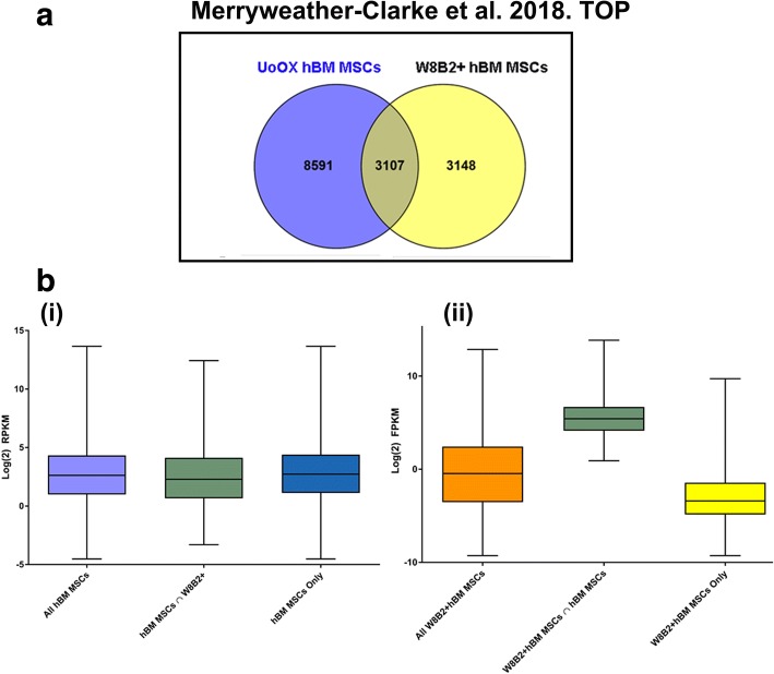
Background: Human bone marrow-derived mesenchymal stem/stromal cells (hBM MSCs) have multiple functions, critical for skeletal formation and function. Their functional heterogeneity, however, represents a major challenge for their isolation and in developing potency and release assays to predict their functionality prior to transplantation. Additionally, potency, biomarker profiles and defining mechanisms of action in a particular clinical setting are increasing requirements of Regulatory Agencies for release of hBM MSCs as Advanced Therapy Medicinal Products for cellular therapies. Since the healing of bone fractures depends on the coupling of new blood vessel formation with osteogenesis, we hypothesised that a correlation between the osteogenic and vascular supportive potential of individual hBM MSC-derived CFU-F (colony forming unit-fibroblastoid) clones might exist.

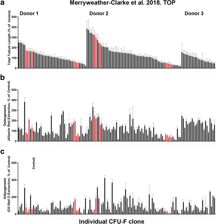
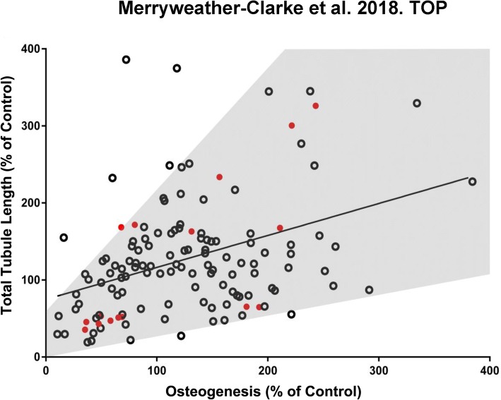
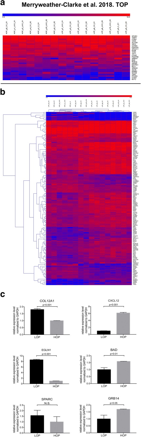
Methods: We tested this by assessing the lineage (i.e. adipogenic (A), osteogenic (O) and/or chondrogenic (C)) potential of individual hBM MSC-derived CFU-F clones and determining if their osteogenic (O) potential correlated with their vascular supportive profile in vitro using lineage differentiation assays, endothelial-hBM MSC vascular co-culture assays and transcriptomic (RNAseq) analyses.

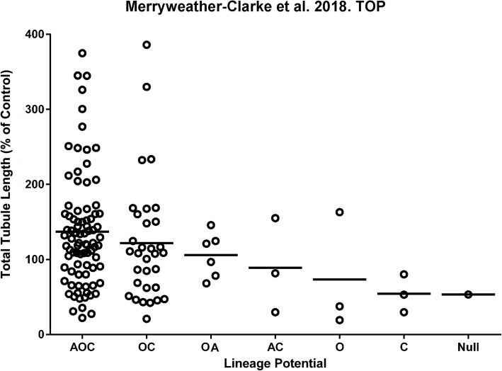
Results: Our results demonstrate that the majority of CFU-F (95%) possessed tri-lineage, bi-lineage or uni-lineage osteogenic capacity, with 64% of the CFU-F exhibiting tri-lineage AOC potential. We found a correlation between the osteogenic and vascular tubule supportive activity of CFU-F clones, with the strength of this association being donor dependent. RNAseq of individual clones defined gene fingerprints relevant to this correlation.

Conclusions: This study identified a donor-dependent correlation between osteogenic and vascular supportive potential of hBM MSCs and important gene signatures that support these functions that are relevant to their bone regenerative properties.

Keywords: CFU-F; Clonal analysis; Mesenchymal stem/stromal cells; Osteogenesis; RNAseq; Vascular supportive capacity.


Conflict of interest statement

Ethics approval and consent to participate

Not applicable.

Consent for publication

Not applicable.

Competing interests

The authors declare that they have no competing interests.

Publisher’s Note

Springer Nature remains neutral with regard to jurisdictional claims in published maps and institutional affiliations.

Figures

Fig. 1
Fig. 1
The relationship between lineage differentiation potential of CFU-F clones and their vascular tubule supportive capacity. A) Clonal cultures were categorised into groups based on their adipogenic (A), osteogenic (O) and chondrogenic (C) differentiation potential and this potency plotted against their ability to support day 14 vascular tubule formation in co-culture assays with HUVEC as measured by the total tubule length. The classification included tri-lineage (AOC), bi-lineage (OC, OA, AC), uni-lineage (O, C) and nullipotent (Null) clones. The total tubule length was normalised as a percentage of that obtained using a control non CFU-F selected hBM MSC sample (Control) which was set at 100%. Three bone marrow aspirates were used to generate 133 CFU-F clones. The bars represent the mean total tubule length (TTL) for each lineage subgroup. Quartiles for TTL are 0 to 68.67%, 68.67 to 116.6%, 116.6 to 156.3% and > 156.3%
Fig. 2
Fig. 2
Quantification of vascular tubule supportive capacity, osteogenesis, and adipogenesis of hBM MSC clonal cultures. Clonal CFU-F cultures of hBM MSCs were expanded into T25 flasks before being assayed quantitatively at P1 for their osteogenic or adipogenic differentiation potential and their ability to support day 14 vascular tubule formation in co-culture assays with HUVEC as measured by the total tubule length. Three donor bone marrows were used (donor 1, donor 2 and donor 3). Data for individual CFU-F clones grouped by donor are shown. a HUVECs were seeded onto hBM MSC monolayers and cultured for 2 weeks before fixation and CD31 antibody staining. Total tubule length was calculated and normalised to the tubule length of a control hBM MSC sample (D), which was run for each separate experiment. The clonal cultures were assayed for their b osteogenic and c adipogenic differentiation potential by 2–3 weeks culture in differentiation media, relative to the control non CFU-F selected hBM MSC sample (Control). This control was set at 100% and all other values normalised against this. Values are mean ± SD of n = 3 replicate cultures. The histograms highlighted in red were used for cell sorting and RNAseq analyses
Fig. 3
Fig. 3
Correlations between osteogenic lineage differentiation potential and vascular tubule supportive capacity. Clonal hBM MSC CFU-F cultures at p1 were assayed quantitatively for their osteogenic differentiation potential after culture in osteogenic differentiation media, relative to the control non CFU-F selected hBM MSC sample (Control), which was set at 100%. and the correlation between osteogenic and vascular supportive activity assessed. Pearson’s correlation coefficient (r) was calculated for all three bone marrow donor aspirates. The red circles are CFU-F clones that were used for cell sorting and RNAseq analyses. There was a moderate positive relationship between the vascular tubule supportive function and the osteogenic potential for all CFU-F clones when these were assessed quantitatively (p [two tailed] < 0.0001; n = 133 clones)
Fig. 4
Fig. 4
Heatmap of transcripts from RNA sequencing CFU-F clones with high versus low osteogenic differentiation capacity and qPCR analyses of candidate genes. a Well characterised hBM MSC genes detected by RNAseq. Expression level is indicated by the scale bar, as counts per million (CPM) mapped reads. Names of sequenced clones indicate whether they exhibited high or low osteogenic potential (HOP or LOP, respectively), good or poor vascular support (g or p), lineage potential (adipogenic (A), Osteogenic (O) and/or chondrogenic (C), and whether they originated from donor 1 (D1) or donor 2 (D2). b Hierarchical clustering of the 16 clones using 161 genes that were differentially expressed between HOP and LOP clones, determined using an FDR < 0.05. c qPCR analyses of COL12A1, EGLN1, CXCL12, BAD and GRB14 genes showing their statistically significant (< 0.05) up- or downregulaton in HOP versus LOP clones. SPARC represented a gene expressed in but not significantly differentially (N.S.) expressed between HOP and LOP clones. Details are described in the “Methods” section
Fig. 5
Fig. 5
Comparison of hBM MSC RNA sequencing results with W8B2+ hBM MSC dataset. a Venn diagram showing the number of expressed genes shared between the datasets from this study (UoOX hBM MSCs) and from the published W8B2+ hBM MSCs data of Zhang et al. [68]. b Boxplots showing the expression levels of all genes, shared genes and unique genes in each dataset. b (i) gene expression levels in hBM MSCs from this study (UoOx hBM MSCs). b (ii) gene expression levels in W8B2+ hBM MSCs from the dataset of Zhang et al. [68]

References

    1. Bianco P, Robey PG. Skeletal stem cells. Development. 2015;142(6):1023–1027. doi: 10.1242/dev.102210. - DOI - PMC - PubMed
    1. Boulais PE, Frenette PS. Making sense of hematopoietic stem cell niches. Blood. 2015;125(17):2621–2629. doi: 10.1182/blood-2014-09-570192. - DOI - PMC - PubMed
    1. Fierro FA, Nolta JA, Adamopoulos IE. Concise review: stem cells in osteoimmunology. Stem Cells. 2017;35(6):1461–1467. doi: 10.1002/stem.2625. - DOI - PMC - PubMed
    1. Kfoury Y, Scadden DT. Mesenchymal cell contributions to the stem cell niche. Cell Stem Cell. 2015;16(3):239–253. doi: 10.1016/j.stem.2015.02.019. - DOI - PubMed
    1. Le Blanc K, Davies LC. MSCs-cells with many sides. Cytotherapy. 2018;20(3):273–278. doi: 10.1016/j.jcyt.2018.01.009. - DOI - PubMed

Publication types