. 2019 Mar;90(3):272-283.
doi: 10.1136/jnnp-2018-318854. Epub 2018 Dec 19.

Platelet abnormalities in Huntington's disease

Affiliations

Platelet abnormalities in Huntington's disease

Hélèna L Denis et al. J Neurol Neurosurg Psychiatry. 2019 Mar.

Abstract

Huntington's disease (HD) is a hereditary disorder that typically manifests in adulthood with a combination of motor, cognitive and psychiatric problems. The pathology is caused by a mutation in the huntingtin gene which results in the production of an abnormal protein, mutant huntingtin (mHtt). This protein is ubiquitously expressed and known to confer toxicity to multiple cell types. We have recently reported that HD brains are also characterised by vascular abnormalities, which include changes in blood vessel density/diameter as well as increased blood-brain barrier (BBB) leakage.

Objectives: Seeking to elucidate the origin of these vascular and BBB abnormalities, we studied platelets that are known to play a role in maintaining the integrity of the vasculature and thrombotic pathways linked to this, given they surprisingly contain the highest concentration of mHtt of all blood cells.

Methods: We assessed the functional status of platelets by performing ELISA, western blot and RNA sequencing in a cohort of 71 patients and 68 age- and sex-matched healthy control subjects. We further performed haemostasis and platelet depletion tests in the R6/2 HD mouse model.

Results: Our findings indicate that the platelets in HD are dysfunctional with respect to the release of angiogenic factors and functions including thrombosis, angiogenesis and vascular haemostasis.

Conclusion: Taken together, our results provide a better understanding for the impact of mHtt on platelet function.


Conflict of interest statement

Competing interests: None declared.

Figures

Figure 1
Figure 1
Roles of platelets. Platelets are primarily involved in (1) angiogenesis, (2) haemostasis and (3) inflammation—all of which can contribute to vascular permeability.
Figure 2
Figure 2
(A) Representative western blot analyses of Htt (mab2166) and anti-polyQ (mab1574) in blood cells. mHtt protein is enriched in platelets compared with blood leucocytes and RBC in patients with HD. PBMC and RBC (hCTRL, n=6; pre-HD, n=6; stage 1–2, n=6; stage 3–4, n=6). (B) Numbers of platelets in hCTRL, pre-HD and patients with HD showed no statistical difference between groups (hCTRL, n=54; pre-HD, n=10; HD, n=50). RNA sequencing of platelets from patients with HD and healthy control group was performed and (C) annotation clustering and heatmap analysis of correlations between disease state and the abundance of the variable RNA (hCTRL, n=5; pre-HD, n=2; HD, n=2). Cold and hot colours represent low and high correlation levels, respectively. Seven RNAs significantly varied between healthy control group and patients with HD and these differences were confirmed by reverse transcription PCR (hCTRL, n=13; pre-HD, n=5; stage 1–2, n=5; stage 3–4, n=2;) (D). Statistical analyses: (A) two-way analysis of variance (ANOVA) followed by Tukey’s multiple comparisons test (**p<0.01, ****p<0.0001); (B) one-way ANOVA, (C.) Kruskal-Wallis with Dunn’s multiple comparison test (*p<0.05, SLC25A43, power statistic=0.71; SLC25A39, power statistic=0.97; SLC25A39, power statistic=0.80; HBG1, power statistic=0.72; HBG2, power statistic=0.95; HBA1, power statistic=0.42; HBA2, power statistic=0.31). HBA1/2, haemoglobin subunit alpha 1/2; HBG1/2, haemoglobin subunit gamma 1/2; hCTRL, healthy control group; Htt, Huntingtin; HD, Huntington’s disease; PBMC, peripheral blood mononuclear cell (lymphocytes and monocytes); pre-HD, premanifest; RBC, red blood cells; RNA, ribonucleic acid:SLC25A43-39-37, solute carrier family 25 member 43-39-37.
Figure 3
Figure 3
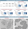
(A) Representative western blot analyses of polyQ (mab1574) and total Htt (mab2166) that were quantified in the pellet of resting platelets (B) as well as platelets activated with 5 µg/mL collagen (C) or 0.5 U/mL thrombin (D) (hCTRL, n=8; pre-HD, n=8; stage 1–2, n=7; stage 3–4, n=7). (E) Representative transmission electron microscopy images of mHtt aggregates using EM48 antibody (1:500, Millipore Sigma: mab5374) as detected in platelets of zQ175 mice. (F) Photomicrograph of the control condition in which platelets from zQ175 mice underwent identical staining but with the omission of the primary antibody. Scale bar= 250 nm. Statistical analyses: Kruskal-Wallis with Dunn’s multiple comparison (*p<0.05, ***p<0.0001) and Wilcoxon signed rank test with theoretical median set at 1 (#p<0.05). hCTRL, healthy control group; HD, Huntington’s disease; Htt, Huntingtin; mHtt, mutant Htt; OCS, open-canalicular system; pre-HD, pre-manifest; α, alpha granule.
Figure 4
Figure 4
ELISA quantified mediators of angiogenesis in (A) purified platelets and (B) media after activation of 108 platelet/mL with collagen or thrombin compared to the resting state. Platelets and media from different HD stage patients were compared with pre-HD and hCTRL (stage 1, n=5; stage 2, n=5; stage 3, n=5; stage 4, n=4; pre-HD, n=5; hCTRL, n=6). The release of both (C) serotonin and (D) PF4 was quantified by ELISA in the activated-platelet media from different HD stage patients, pre-HD and hCTRL (stage 2, n=4; stage 4, n=4; pre-HD, n=8; hCTRL, n=9). Statistical analyses: Kruskal-Wallis with Dunn’s multiple comparison test (*p<0.05, **p<0.01) and Wilcoxon signed rank test with theoretical median set at 1 (#p<0.05). ANG-2, angiopoietin-2; FGF basic, basic fibroblast growth factor; hCTRL, healthy control group; HD, Huntington’s disease; HGF, hepatocyte growth factor; IL-8, interleukin 8; PDGFR, platelet-derived growth factor; TIMP-1, metalloproteinase inhibitor 1; TIMP-2, metalloproteinase inhibitor 2; TNFα, tumour necrosis factor; pre-HD, pre-manifest.
Figure 5
Figure 5
(A) Tail bleeding test revealed a significant decrease in bleeding time and blood volume in old (12/13 weeks) R6/2 mice compared with WT mice (bleeding time: 4–6 weeks, WT=6, R6/2=8; 8–9 weeks, WT=8, R6/2=9; 12–13 weeks, WT=15, R6/2=13; lost blood volume: 4–6 weeks, WT=6, R6/2=8; 8–9 weeks, WT=9, R6/2=7; 12–13 weeks, WT=9, R6/2=8). (B) H&E staining and quantification of blood vessels in 12-week-old R6/2 mice showed a significant decrease of vessel density compared with 12-week-old WT mice (R6/2, n=4; WT, n=4). Statistical analysis: two-way analysis of variance followed by Sidak multiple comparison test (A) and Mann-Whitney test (*p<0.05) (B). B, bone; h, hair; H&E, hematoxylin eosin; m, muscle; v, vessel, WT, wild type.
Figure 6
Figure 6
(A) Timeline of platelet depletion protocol with antiplatelet antibody (R-300) or non-immune antibodies (C-301) administered to 9-week-old WT or R6/2 mice. (B) Illustration of depletion protocol. (C) Number of platelets in undepleted (+) or depleted (−) WT and R6/2 mice (+WT, n=5; −WT, n=5; +R6/2, n=10; −R6/2, n=10). Platelet depletion was successful given that no platelets were found in depleted mice. (D) Open field test results indicated that there was no significant difference between depleted or undepleted mice. (E) Permeability of BBB using EB injection before sacrifice of mice demonstrated a significant increase of permeability in the cerebellum of undepleted R6/2 mice compared with WT and depleted R6/2 mice. No difference was observed in the cortex (data not shown). (F) Western blot analysis showed a decrease of claudin-5, a tight junction protein involved on BBB homeostasis, in undepleted R6/2 striatum that was normalised by platelet depletion. Statistical analyses: Kruskal-Wallis test followed by Dunn’s multiple comparison test (*p<0.05, **p<0.01). BBB, blood–brain barrier; EB, Evans blue; Plts, platelets, WT, wild type.

References

    1. Bates GP, Dorsey R, Gusella JF. Huntington disease. Nat Rev Dis Primer 2015;1:nrdp20155. - PubMed
    1. Sassone J, Colciago C, Cislaghi G, et al. . Huntington's disease: the current state of research with peripheral tissues. Exp Neurol 2009;219:385–97. 10.1016/j.expneurol.2009.05.012 - DOI - PubMed
    1. Weiss A, Träger U, Wild EJ, et al. . Mutant huntingtin fragmentation in immune cells tracks Huntington's disease progression. J Clin Invest 2012;122:3731–6. 10.1172/JCI64565 - DOI - PMC - PubMed
    1. Drouin-Ouellet J, Sawiak SJ, Cisbani G, et al. . Cerebrovascular and blood-brain barrier impairments in Huntington's disease: potential implications for its pathophysiology. Ann Neurol 2015;78:160–77. 10.1002/ana.24406 - DOI - PubMed
    1. Hsiao HY, Chen YC, Huang CH, et al. . Aberrant astrocytes impair vascular reactivity in Huntington disease. Ann Neurol 2015;78:178–92. 10.1002/ana.24428 - DOI - PubMed

Publication types