. 2019 Feb 22;294(8):2903-2912.
doi: 10.1074/jbc.RA118.006237. Epub 2018 Dec 19.

FAD-sequestering proteins protect mycobacteria against hypoxic and oxidative stress

Affiliations

FAD-sequestering proteins protect mycobacteria against hypoxic and oxidative stress

Liam K Harold et al. J Biol Chem. .

Abstract

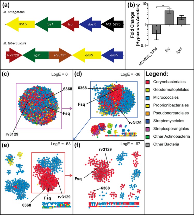
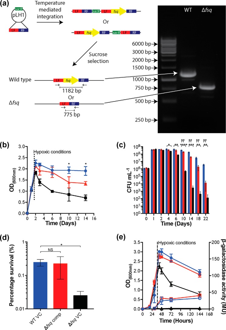
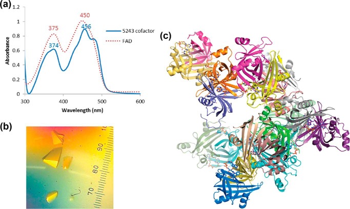
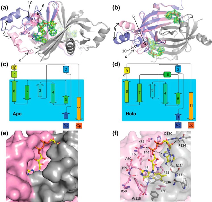
The ability to persist in the absence of growth triggered by low oxygen levels is a critical process for the survival of mycobacterial species in many environmental niches. MSMEG_5243 (fsq), a gene of unknown function in Mycobacterium smegmatis, is up-regulated in response to hypoxia and regulated by DosRDosS/DosT, an oxygen- and redox-sensing two-component system that is highly conserved in mycobacteria. In this communication, we demonstrate that MSMEG_5243 is a flavin-sequestering protein and henceforth refer to it as Fsq. Using an array of biochemical and structural analyses, we show that Fsq is a member of the diverse superfamily of flavin- and deazaflavin-dependent oxidoreductases (FDORs) and is widely distributed in mycobacterial species. We created a markerless deletion mutant of fsq and demonstrate that fsq is required for cell survival during hypoxia. Using fsq deletion and overexpression, we found that fsq enhances cellular resistance to hydrogen peroxide treatment. The X-ray crystal structure of Fsq, solved to 2.7 Å, revealed a homodimeric organization with FAD bound noncovalently. The Fsq structure also uncovered no potential substrate-binding cavities, as the FAD is fully enclosed, and electrochemical studies indicated that the Fsq:FAD complex is relatively inert and does not share common properties with electron-transfer proteins. Taken together, our results suggest that Fsq reduces the formation of reactive oxygen species (ROS) by sequestering free FAD during recovery from hypoxia, thereby protecting the cofactor from undergoing autoxidation to produce ROS. This finding represents a new paradigm in mycobacterial adaptation to hypoxia.

Keywords: reactive oxygen species (ROS); mycobacteria; Mycobacterium smegmatis; hypoxia; flavin; flavin adenine dinucleotide (FAD); bacterial genetics; oxidative stress; anoxia; dos regulon; DosR; oxidoreductase.


Conflict of interest statement

The authors declare that they have no conflicts of interest with the contents of this article

Figures

Figure 1.
Figure 1.
Organization, regulation, and abundance of Fsq. a, genomic context of fsq in M. smegmatis and its homolog rv3129 in M. tuberculosis. Matching colors between M. smegmatis and M. tuberculosis represent homologous genes. Gene sizes are not to scale. b, comparison of expression ratio of MSMEG_6368 and fsq, with tgs1 as a positive control, from cells harvested at 6 days' growth in aerobic and hypoxic conditions. The gene sigA was used as the reference. Error bars represent S.D. of biological triplicates. Statistical testing was performed using an unpaired Student's t test, where ** = p < 0.01. c–f, SSNs of Fsq-related proteins are shown with different cutoffs. In an SSN, protein sequences are represented as nodes (dots) that are joined by edges (lines) which represent a similarity metric such as Basic Local Alignment Search Tool (BLAST) E-value (48). Nodes are colored according to taxonomic order. c, SSN with no logE cutoff applied. d, SSN of Fsq-related proteins with a logE cutoff of −36. A large heterogeneous cluster containing Fsq, MSMEG_6368, and rv3129 separates from several small clusters. e, the large heterogenous cluster examined with a cutoff of −53. A large compact cluster of mycobacterial sequences separates from other clusters. f, the large cluster from previous panel with a cutoff of −67. A cluster containing MSMEG_6368 separates from one containing Fsq and rv3129, suggesting that they may have distinct functions.
Figure 2.
Figure 2.
Creation and hypoxic survival of Δfsq markerless deletion mutant. a, diagram of fsq markerless deletion construction. b and c, long-term survival of M. smegmatis WT (blue) compared with ΔdosR (black) and Δfsq (red) mutants under hypoxic conditions. Growth was in HdB minimal medium supplemented with 22 mm glycerol. Entry of the vials into a hypoxic state was estimated by the decolorization of methylene blue (1.5 μg/ml) which occurred at 27 h. b, growth measured using optical density at 600 nm wavelength with an average of three biological replicates shown with error bars representing S.D. Unpaired Student's t test of WT compared with Δfsq. *, p < 0.05 at each time point. c, survival measured using colony-forming units (CFU) with an average of three biological replicates shown with error bars representing S.D. Unpaired Student's t test of WT compared with Δfsq and WT compared with ΔdosR at each time point. *, p < 0.05; **, p < 0.01; ***, p < 0.001; ****, p < 0.0001. d, complementation of survival defect of Δfsq mutant using pLH2 (fsq+) (Δfsq comp) (red) compared with an empty vector control in wild type (WT VC) (blue) and an empty vector in Δfsq mutant (Δfsq VC) (black). Vectors were induced using 20 ng/ml tetracycline just prior to entry into hypoxia. Survival percentage of day 18 with an average of three biological replicates shown with error bars representing S.D. Unpaired Student's t test of WT compared with Δfsq comp and WT compared with Δfsq VC. *, p < 0.05. e, measurement of DosR activity through hyd3 LacZ expression in M. smegmatis WT compared with ΔdosR and Δfsq mutants under hypoxic conditions. Strains were grown into hypoxia in HdB with 22 mm glycerol. Entry into hypoxia was indicated by methylene blue (1.5 μg/ml) control vials. Samples (2 ml) were taken at 27, 48, 72, and 144 h and used to preform β-gal assays to assess the level of dosR activity. Key: WT, blue circles; ΔdosR mutant, black triangles; Δfsq mutant, red squares; β-gal activity, open symbols; optical density (600 nm), closed symbols. Each sample shows average of a biological triplicate with error bars representing S.D.
Figure 3.
Figure 3.
a, UV-visible absorbance spectra comparing the cofactor co-purified with Fsq (blue) to FAD (red). b, crystals of Fsq bound to the co-purified FAD, with no cofactor stripping or additional reconstitution (40× magnification). c, the 20 chains of apo- and holo-Fsq found in the asymmetric unit. 17 chains were bound to FAD.
Figure 4.
Figure 4.
Structure of apo- and holo-FAD Fsq. a and b, side (a) and top (b) views of the overlaid structures of apo (blue, chain P) and holo (pink, chain B) Fsq. The secondary chain of the dimer for both structures is shown in gray. FAD bound to the holo-structure is shown in yellow and the electron density for the cofactor on both binding sites of the dimer is shown, representing the Fo − Fc omit map contoured at 3 σ. Key loops and α-helixes are labeled to reference parts c and d. c and d, topology maps of the apo- and holo-structures, respectively, adapted from the cartoons generated by Pro-origami. e, solvent-accessible surface of Fsq showing the buried flavin moiety in the holo-structure. f, residues involved in FAD binding.
Figure 5.
Figure 5.
Electrochemical properties of Fsq. a, cyclic voltammograms of Fsq (blue) and FAD (red) adsorbed onto modified glassy carbon electrodes. Scans were conducted at 50 mV/s in the direction indicated by the arrows in 100 mm sodium phosphate, pH 7.3. Oxidative and reductive peaks are labeled. The cyclic voltammogram of Fsq is scaled by a factor of 7.5 for comparison. b, effect of pH on the redox potential of Fsq and FAD. Over the range tested Fsq and FAD varied by −45 mV/pH unit and −39 mV/pH unit, respectively. NHE, normal hydrogen electrode; E, electrochemical potential.
Figure 6.
Figure 6.
Fsq heightens resistance to hydrogen peroxide. a, challenge of WT compared with Δfsq with 20 or 50 mm hydrogen peroxide for 3 h. Cells were grown aerobically in HdB with 22 mm glycerol for 10 days before harvest and resuspension in phosphate buffer saline with 0.5% tyloxapol. Cells were normalized to 1 optical density (600 nm) before treatment. b, challenge of WT compared with Δfsq with 20 or 50 mm hydrogen peroxide for 3 h. Cells were grown hypoxically in HdB with 22 mm glycerol for 10 days before harvest and resuspension in phosphate buffer saline with 0.5% tyloxapol. Cells were normalized to 1 optical density (600 nm) before treatment. c, challenge of WT either expressing fsq (fsq+) (WT pLH2) or a vector control (VC) (WT pMind) control induced with 20 ng−1 ml tetracycline, challenged with 50 or 75 mm hydrogen peroxide for 3 h. Cells were grown aerobically to exponential phase n HdB with 22 mm glycerol before harvest and resuspension in phosphate buffer saline with 0.5% tyloxapol. Cells were normalized to 1 optical density (600 nm) before treatment. a–c, error bars represent S.D. of three biological replicates. Unpaired Student's t test compared with WT for each treatment, where *, p < 0.05; **, p < 0.01.

References

    1. Miller D. M., Buettner G. R., and Aust S. D. (1990) Transition-metals as catalysts of autoxidation reactions. Free Radic. Biol. Med. 8, 95–108 10.1016/0891-5849(90)90148-C - DOI - PubMed
    1. Massey V. (1994) Activation of molecular-oxygen by flavins and flavoproteins. J. Biol. Chem. 269, 22459–22462 - PubMed
    1. Piddington D. L., Fang F. C., Laessig T., Cooper A. M., Orme I. M., and Buchmeier N. A. (2001) Cu,Zn superoxide dismutase of Mycobacterium tuberculosis contributes to survival in activated macrophages that are generating an oxidative burst. Infect. Immun. 69, 4980–4987 10.1128/IAI.69.8.4980-4987.2001 - DOI - PMC - PubMed
    1. Ng V. H., Cox J. S., Sousa A. O., MacMicking J. D., and McKinney J. D. (2004) Role of KatG catalase-peroxidase in mycobacterial pathogenesis: Countering the phagocyte oxidative burst. Mol. Microbiol. 52, 1291–1302 10.1111/j.1365-2958.2004.04078.x - DOI - PubMed
    1. Flynn J. L., and Chan J. (2001) Immunology of tuberculosis. Annu. Rev. Immunol. 19, 93–129 10.1146/annurev.immunol.19.1.93 - DOI - PubMed

Publication types

LinkOut - more resources