. 2019 Mar 5;93(6):e02011-18.
doi: 10.1128/JVI.02011-18. Print 2019 Mar 15.

Flexibility In Vitro of Amino Acid 226 in the Receptor-Binding Site of an H9 Subtype Influenza A Virus and Its Effect In Vivo on Virus Replication, Tropism, and Transmission

Affiliations

Flexibility In Vitro of Amino Acid 226 in the Receptor-Binding Site of an H9 Subtype Influenza A Virus and Its Effect In Vivo on Virus Replication, Tropism, and Transmission

Adebimpe O Obadan et al. J Virol. .

Abstract

Influenza A viruses (IAVs) remain a significant public health threat, causing more than 300,000 hospitalizations in the United States during the 2015-2016 season alone. While only a few IAVs of avian origin have been associated with human infections, the ability of these viruses to cause zoonotic infections further increases the public health risk of influenza. Of these, H9N2 viruses in Asia are of particular importance as they have contributed internal gene segments to other emerging zoonotic IAVs. Notably, recent H9N2 viruses have acquired molecular markers that allow for a transition from avian-like to human-like terminal sialic acid (SA) receptor recognition via a single amino acid change at position 226 (H3 numbering), from glutamine (Q226) to leucine (L226), within the hemagglutinin (HA) receptor-binding site (RBS). We sought to determine the plasticity of amino acid 226 and the biological effects of alternative amino acids on variant viruses. We created a library of viruses with the potential of having any of the 20 amino acids at position 226 on a prototypic H9 HA subtype IAV. We isolated H9 viruses that carried naturally occurring amino acids, variants found in other subtypes, and variants not found in any subtype at position 226. Fitness studies in quails revealed that some natural amino acids conferred an in vivo replication advantage. This study shows the flexibility of position 226 of the HA of H9 influenza viruses and the resulting effect of single amino acid changes on the phenotype of variants in vivo and in vitroIMPORTANCE A single amino acid change at position 226 in the hemagglutinin (HA) from glutamine (Q) to leucine (L) has been shown to play a key role in receptor specificity switching in various influenza virus HA subtypes, including H9. We tested the flexibility of amino acid usage and determined the effects of such changes. The results reveal that amino acids other than L226 and Q226 are well tolerated and that some amino acids allow for the recognition of both avian and human influenza virus receptors in the absence of other changes. Our results can inform better avian influenza virus surveillance efforts as well as contribute to rational vaccine design and improve structural molecular dynamics algorithms.

Keywords: H9 subtype; avian viruses; evolution; quail; receptor; sialic acid; transmission; zoonosis.


Figures

FIG 1
FIG 1
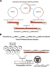
Schematic overview of the steps to generate the degenerate H9 HA PCR product and rescue the H9 HA virus library. (A) The wild-type WF10 HA plasmid was split into 2 plasmids with designed primers (pDPAO1-767 and pDPAO738-1742). The HA PCR product (1 to 767) carrying the mouse RNA polymerase terminator sequence and the degenerate NNN codon at position 226 was generated from the pDPAO1-767 plasmid either with a specific primer with the NNN codon (nnn226) or with a mix of primers able to introduce all 20 amino acids (equi226). Another PCR product with the remaining HA (residues 738 to 1742) and human polymerase 1 promoter was generated from pDPAO738-1742. Using overlapping PCR, a full-length HA was obtained with the degenerate codon at position 226 flanked by the mouse RNA polymerase terminator sequence and the human pol 1 promoter. (B) Generation of virus library by PCR-based reverse genetics using the 226HA PCR product and 7 plasmids carrying proteins from WF10 (H9N2) or PR8 (H1N1).
FIG 2
FIG 2
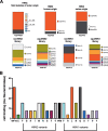
Amino acid diversity at position 226 and impact of mutations on receptor avidity. (A) The amino acid present at position 226 in natural isolates of H9Nx viruses by species of origin (avian, human, and swine) compared to the amino acid identified experimentally using either the nnn226 approach or the equi226 approach on an H9N1 or an H9N2 backbone. (B) Receptor-binding avidity of viruses using chicken red blood cells treated with increasing concentrations of neuraminidase from Clostridium perfringens and comparison to that of wild-type WF10 (L226) virus and wild-type PR8 virus.
FIG 3
FIG 3
In vitro replication of H9N2 and H9N1 variants. Replication of H9N2 and H9N1 viruses in mammalian MDCK cells (A, C) and avian-origin DF1 cells (B, D) at 37°C. Naturally occurring amino acids previously found in H9 HA are shown as squares; others are shown as triangles. Confluent monolayers of MDCK or DF1 cells were infected with viruses at an MOI of 0.01, and supernatant was collected at 0, 6, 12, 24, 48, and 72 hpi. The virus in supernatant collected from MDCK cells was quantified by determination of the TCID50 using the Reed and Muench method (62). The plotted data represent means ± standard errors. WT, wild type.
FIG 4
FIG 4
The sialic acid specificity of viruses is dependent on the amino acid at position 226 in glycan array assays. The receptor-binding specificity of a subset of mutant viruses in the H9N2 backbone was determined using a glycan array. L, Q, M, F, I, S, H, N, V, C, T, and G correspond to amino acid mutations at position 226 in the HA of H9N2 viruses. Glycans on the array comprise nonsialoside controls (glycans 1 to 10; gray), α2-3 sialosides (glycans 11 to 79; yellow), and α2-6 sialosides (glycans 80 to 135; green). Glycans are grouped by structure type: L, linear; O, O linked; N, N linked; and Lx, sialyl-Lewis X. RFU, relative fluorescent units. Plotted data represent means ± standard errors (SEM).
FIG 5
FIG 5
The sialic acid specificity of viruses is dependent on the amino acid at position 226 in solid-phase binding assays. The receptor-binding specificity of mutant viruses on the H9N2 backbone was determined using various concentrations of sialic acid conjugated to biotinylated sialylglycopolymers (3SLN and 6SLN) via direct solid-phase binding assays. The y axis represents absorbance (optical density) values at 450 nm. The x axis corresponds to the concentrations of serially diluted 3SLN or 6SLN sialylglycopolymers. As expected, the H9N2 L226 virus bound mainly 6SLN-PAA-biotin while the Q226, C226, H226, S226, and T226 viruses bound more to the 3SLN-PAA-biotin moiety. The F226, G226, and I226 viruses showed a preference for both 3SLN and 6SLN sugars. The figure represents the results of a prototypical assay with samples run in duplicate.
FIG 6
FIG 6
Pattern of virus attachment to quail tracheal tissues. The attachment patterns of the var viruses to quail tracheal tissue sections were determined. Virus attachment was seen as red staining on the tracheal surface epithelium. The intensity of binding to the surface epithelium of the trachea was scored as none (−), low (+), moderate (++), and intense (+++) (Table 2). Representative bright-field images are shown.
FIG 7
FIG 7
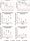
Replication and transmission phenotype of mutant viruses in quails. Quails were randomly divided into five groups; one group (inoculated with PBS) served as a control (not shown). At 4 to 5 weeks of age, 6 birds were inoculated intranasally, intratracheally, and cloacally with a mixture of viruses with or without Q226 and/or L226 at 106 TCID50/ml per bird. Group 1 received only H9N2 variant viruses without L226 and Q226 (varΔLQ), group 2 received variant viruses including the Q226 virus (var+Q), group 3 received variant viruses including the L226 virus (var+L), and the birds in group 4 received all variant viruses, including the Q226 and L226 viruses (var+LQ). On day 1 postinfection, 6 naive quails were introduced as direct-contact animals. (A, C, E) Viral shedding was quantified by quantitative reverse transcriptase PCR in tracheal swabs at 3, 5, 7, and 9 dpi and in tracheal and lung homogenates at 5 dpi for inoculated birds. (B, D, F) Viral shedding was quantified by quantitative reverse transcriptase PCR in tracheal swabs at 2, 4, 6, and 8 dpc and in tracheal and lung homogenates at 6 dpc for direct-contact birds. Statistically significant differences between the varΔLQ group compared to the other groups are indicated. **, P < 0.001; *, P < 0.05.
FIG 8
FIG 8
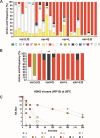
Frequency of amino acids present in tracheal swab samples of inoculated and contact quails and temperature stability of H9N2 variant viruses. The viruses in tracheal swab specimens collected at 3 dpi from inoculated quails (A) and 8 dpc from contact quails (B) were sequenced by NGS, and the amino acid frequency was analyzed using Geneious (version 10.2.3) software. Each bar represents a quail in the respective group, and each color represents a variant virus. Single asterisks refer to variant viruses included in the virus mix and not identified in tracheal swabs, while the double asterisk shows the K226 variant virus not included in the virus mix yet identified in contact quail. (C) Temperature stability of H9N2 variant viruses at 56°C. Variant viruses diluted to 128 HAU/50 μl were incubated at 56°C. Samples collected at 0, 15, 30, 60, 120, 180, and 240 min postincubation were used for hemagglutination assays. Treatment was carried out in quadruplicate. L, Q, M, F, I, S, H, N, V, C, T, and G correspond to the amino acid mutations at position 226 in the HA of H9N2 viruses. Naturally occurring amino acids previously found in H9 HA are shown as squares; others are shown as triangles. Differences were observed depending on which amino acid was present at position 226. By 1 h at 56°C, all H9N2 viruses had hemagglutination titers (HA titers) of less than 2 HAU, except viruses with aliphatic amino acids, V226, L226, and I226 viruses, as well as the C226 and Q226 viruses. After 4 h, only the viruses with V226 and L226 showed any hemagglutination titer and were the most stable viruses.

References

    1. de Graaf M, Fouchier RA. 2014. Role of receptor binding specificity in influenza A virus transmission and pathogenesis. EMBO J 33:823–841. doi:10.1002/embj.201387442. - DOI - PMC - PubMed
    1. Weis W, Brown JH, Cusack S, Paulson JC, Skehel JJ, Wiley DC. 1988. Structure of the influenza virus haemagglutinin complexed with its receptor, sialic acid. Nature 333:426–431. doi:10.1038/333426a0. - DOI - PubMed
    1. Skehel JJ, Bayley PM, Brown EB, Martin SR, Waterfield MD, White JM, Wilson IA, Wiley DC. 1982. Changes in the conformation of influenza virus hemagglutinin at the pH optimum of virus-mediated membrane fusion. Proc Natl Acad Sci U S A 79:968–972. doi:10.1073/pnas.79.4.968. - DOI - PMC - PubMed
    1. Garten W, Bosch FX, Linder D, Rott R, Klenk HD. 1981. Proteolytic activation of the influenza virus hemagglutinin: the structure of the cleavage site and the enzymes involved in cleavage. Virology 115:361–374. doi:10.1016/0042-6822(81)90117-3. - DOI - PubMed
    1. Suzuki Y, Ito T, Suzuki T, Holland RE Jr, Chambers TM, Kiso M, Ishida H, Kawaoka Y. 2000. Sialic acid species as a determinant of the host range of influenza A viruses. J Virol 74:11825–11831. doi:10.1128/JVI.74.24.11825-11831.2000. - DOI - PMC - PubMed

Publication types

MeSH terms

LinkOut - more resources