

Review
doi: 10.1128/JVI.01429-17.
Print 2019 Mar 15.
Tetraspanins: Architects of Viral Entry and Exit Platforms
Affiliations
- PMID: 30567993
- PMCID: PMC6401424
- DOI: 10.1128/JVI.01429-17
Item in Clipboard
Review
Tetraspanins: Architects of Viral Entry and Exit Platforms
J Virol.
.
Abstract
Host factors render cells susceptible to viral infection. One family of susceptibility factors, the tetraspanin proteins, facilitate enveloped virus entry by promoting virus-cell membrane fusion. They also facilitate viral egress from infected cells. In this Gem, we discuss recent insights into how tetraspanins assemble viral entry and exit platforms on cell membranes, and we speculate that tetraspanins contribute to nonviral membrane fusions by similar mechanisms.
Keywords: coronavirus; membrane fusion; tetraspanin; virus entry.
Copyright © 2019 American Society for Microbiology.
Figures
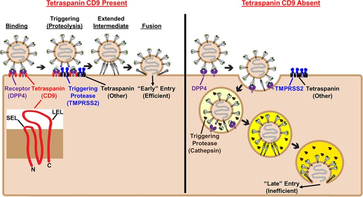
Tetraspanins construct virus entry platforms and dictate CoV entry routes. (Inset) Tetraspanins are composed of four transmembrane spans connected by one large extracellular loop (LEL) and one small extracellular loop (SEL), with short N- and C-terminal tails protruding into the cytosol. (Left) To drive viral entry into host cells, MERS-CoV S (MERS-S) proteins (gray) bind dipeptidyl peptidase 4 (DPP4) receptors (purple) via their receptor binding domains (green). Receptor engagement exposes substrates (blue stars) susceptible to cleavage by cellular proteases, including type II transmembrane protease serine subtype 2 (TMPRSS2) (blue). The tetraspanin CD9 (red) links DPP4 to TMPRSS2 on the plasma membrane. DPP4-TMPRSS2 linkages facilitate the rapid proteolytic triggering of MERS-S proteins after DPP4 binding. Proteolytically triggered MERS-S relocates its receptor binding domains and unfolds into an extended intermediate structure that embeds hydrophobic fusion peptides into target cell membranes. Refolding of intermediates then pulls virus and cell membranes together to catalyze membrane fusion and “early” entry at or near the cell surface. (Right) In the absence of CD9, DPP4 and TMPRSS2 are not linked, and MERS-S proteins are not efficiently triggered on the cell surface. MERS-CoVs are instead endocytosed and thus encounter endosomal cathepsins (brown). At low pH (yellow), cathepsins cleave MERS-S proteins, triggering inefficient, “late” entry in the endosomal network.
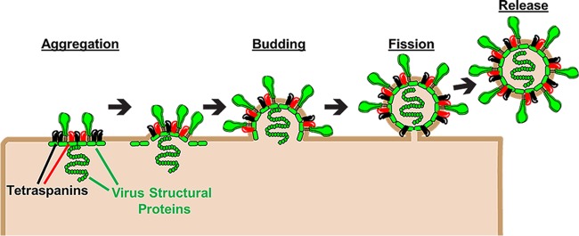
Tetraspanins construct virus exit platforms. Tetraspanins (black and red) associate with viral structural proteins (green), leading to their aggregation into TEMs. Tetraspanins also induce positive membrane curvatures that may promote nascent virus budding. Finally, tetraspanins facilitate membrane fission, allowing for virus release.
References
-
- Huang S, Yuan S, Dong M, Su J, Yu C, Shen Y, Xie X, Yu Y, Yu X, Chen S, Zhang S, Pontarotti P, Xu A. 2005. The phylogenetic analysis of tetraspanins projects the evolution of cell-cell interactions from unicellular to multicellular organisms. Genomics 86:674–684. doi: 10.1016/j.ygeno.2005.08.004. - DOI - PubMed
-
- Yang X, Claas C, Kraeft S-K, Chen LB, Wang Z, Kreidberg JA, Hemler ME. 2002. Palmitoylation of tetraspanin proteins: modulation of CD151 lateral interactions, subcellular distribution, and integrin-dependent cell morphology. Mol Biol Cell 13:767–781. doi: 10.1091/mbc.01-05-0275. - DOI - PMC - PubMed
-
- Charrin S, Manie S, Oualid M, Billard M, Boucheix C, Rubinstein E. 2002. Differential stability of tetraspanin/tetraspanin interactions: role of palmitoylation. FEBS Lett 516:139–144. - PubMed
Publication types
MeSH terms
Substances
LinkOut - more resources
Full Text Sources
Medical
Research Materials
