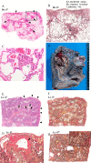
The Analysis of Surgical Lung Biopsy and Explanted Lung Specimens Sheds Light on the Pathological Progression of Chronic Bird-related Hypersensitivity Pneumonitis
- PMID: 30568114
- PMCID: PMC6522403
- DOI: 10.2169/internalmedicine.1142-18
The Analysis of Surgical Lung Biopsy and Explanted Lung Specimens Sheds Light on the Pathological Progression of Chronic Bird-related Hypersensitivity Pneumonitis
Abstract
Chronic hypersensitivity pneumonitis is an interstitial pneumonia caused by an immunological reaction to the chronic inhalation of an antigen. Little is known, however, about the pathological change of the pulmonary lesions. A 33-year-old man was diagnosed with chronic bird-related hypersensitivity pneumonitis based on the findings of a surgical lung biopsy and an inhalation provocation test. He underwent lung transplantation at 8 years after the diagnosis because of disease progression. We conclude that the analysis of the explant suggests that the presence of extensive fibrosis in the centrilobular and perilobular area with bridging fibrosis is a form of pathological progression of chronic hypersensitivity pneumonitis.
Keywords: bridging fibrosis; centrilobular fibrosis; chronic hypersensitivity pneumonitis; lung transplantation; subpleural and paraseptal fibrosis.
Conflict of interest statement
Figures


Similar articles
-
Pathology of hypersensitivity pneumonitis.Curr Opin Pulm Med. 2008 Sep;14(5):440-54. doi: 10.1097/MCP.0b013e3283043dfa. Curr Opin Pulm Med. 2008. PMID: 18664975 Review.
-
Centrilobular Fibrosis in Fibrotic (Chronic) Hypersensitivity Pneumonitis, Usual Interstitial Pneumonia, and Connective Tissue Disease-Associated Interstitial Lung Disease.Arch Pathol Lab Med. 2020 Dec 1;144(12):1509-1516. doi: 10.5858/arpa.2019-0628-RA. Arch Pathol Lab Med. 2020. PMID: 32233994 Review.
-
Chronic hypersensitivity pneumonitis in patients diagnosed with idiopathic pulmonary fibrosis: a prospective case-cohort study.Lancet Respir Med. 2013 Nov;1(9):685-94. doi: 10.1016/S2213-2600(13)70191-7. Epub 2013 Oct 21. Lancet Respir Med. 2013. PMID: 24429272
-
Pathological differentiation of chronic hypersensitivity pneumonitis from idiopathic pulmonary fibrosis/usual interstitial pneumonia.Histopathology. 2012 Dec;61(6):1026-35. doi: 10.1111/j.1365-2559.2012.04322.x. Epub 2012 Aug 8. Histopathology. 2012. PMID: 22882269
-
Can CT distinguish hypersensitivity pneumonitis from idiopathic pulmonary fibrosis?AJR Am J Roentgenol. 1995 Oct;165(4):807-11. doi: 10.2214/ajr.165.4.7676971. AJR Am J Roentgenol. 1995. PMID: 7676971
References
-
- Yoshizawa Y, Ohtani Y, Hayakawa H, Sato A, Suga M, Ando M. Chronic hypersensitivity pneumonitis in Japan: a nationwide epidemiologic survey. J Allergy Clin Immunol 103: 315-320, 1999. - PubMed
-
- Morell F, Villar A, Montero MÁ, et al. . Chronic hypersensitivity pneumonitis in patients diagnosed with idiopathic pulmonary fibrosis: a prospective case-cohort study. Lancet Respir Med 1: 685-694, 2013. - PubMed
-
- Chiba S, Tsuchiya K, Akashi T, et al. . Chronic hypersensitivity pneumonitis with a usual interstitial pneumonia-like pattern: correlation between histopathological and clinical findings. Chest 149: 1473-1481, 2016. - PubMed
-
- Trahan S, Hanak V, Ryu JH, Myers JL. Role of surgical lung biopsy in separating chronic hypersensitivity pneumonia from usual interstitial pneumonia/idiopathic pulmonary fibrosis: analysis of 31 biopsies from 15 patients. Chest 134: 126-132, 2008. - PubMed
-
- Akashi T, Takemura T, Ando N, et al. . Histopathologic analysis of sixteen autopsy cases of chronic hypersensitivity pneumonitis and comparison with idiopathic pulmonary fibrosis/usual interstitial pneumonia. Am J Clin Pathol 131: 405-415, 2009. - PubMed

