. 2019 Mar;33(3):653-661.
doi: 10.1038/s41375-018-0306-7. Epub 2018 Dec 19.

Wnt5a causes ROR1 to complex and activate cortactin to enhance migration of chronic lymphocytic leukemia cells

Affiliations

Wnt5a causes ROR1 to complex and activate cortactin to enhance migration of chronic lymphocytic leukemia cells

Md Kamrul Hasan et al. Leukemia. 2019 Mar.

Abstract

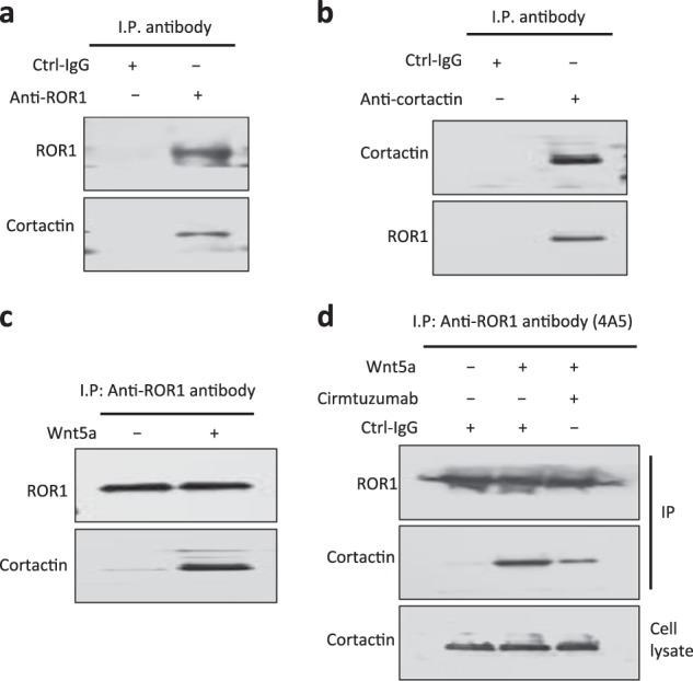
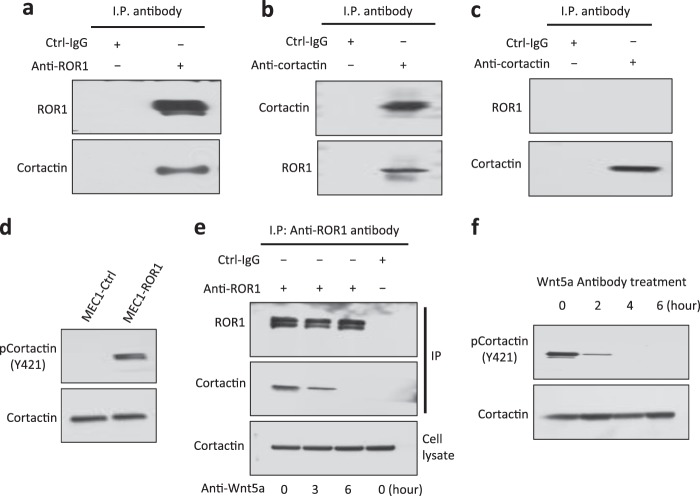
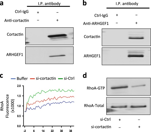
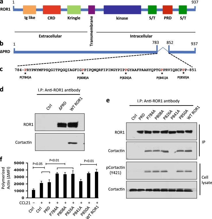
Chronic lymphocytic leukemia cells (CLL) migrate between the blood and lymphoid tissues in response to chemokines. Such migration requires structured cytoskeletal-actin polymerization, which may involve the protein cortactin. We discovered that treatment of CLL cells with Wnt5a causes Receptor tyosin kinase-like orphan receptor 1 (ROR1) to bind cortactin, which undergoes tyrosine phosphorylation at Y421, recruits ARHGEF1, and activates RhoA, thereby enhancing leukemia-cell migration; such effects could be inhibited by cirmtuzumab, a humanized mAb specific for ROR1. We transfected the CLL-cell-line MEC1 with either full-length ROR1 or various mutant forms of ROR1 to examine the structural features required for binding cortactin. We found that the proline-rich domain (PRD) was necessary for ROR1 to recruit cortactin. We generated MEC1 cells that each expressed a mutant form of ROR1 with a single amino-acid substitution of alanine (A) for proline (P) in potential SH3-binding sites in the ROR1-PRD at positions 784, 808, 826, 841, or 850. In contrast to wild-type ROR1, or other ROR1P=>A mutants, ROR1P(841)A failed to complex with cortactin or ARHGEF1 in response to Wnt5a. Moreover, Wnt5a could not induce MEC1-ROR1P(841)A to phosphorylate cortactin or enhance CLL-cell F-actin polymerization. Taken together, these studies show that cortactin plays an important role in ROR1-dependent Wnt5a-enhanced CLL-cell migration.


Conflict of interest statement

The authors declare that they have no conflict of interest.

Figures

Fig. 1
Fig. 1
Phosphorylation of cortactin is high in ROR1Pos CLL. a Immunoblot analysis of lysates prepared from primary ROR1Pos or ROR1Neg CLL cells of different patients; filters were probed with anti-cortactin, anti-phospho-cortactin (anti-pCortactin (Y421)), or anti-ROR1 antibody, as indicated on the left. The numbers above the top lane are ratios of band integrated optical density (IOD) of phosphorylated versus total cortactin. b Phosphorylation of cortactin (at Y421) was assessed by immunoblot analysis of lysates prepared from primary CLL cells of different patients with CLL cells that did (ROR1Pos (n = 13)) or did not (ROR1Neg (n = 11)) express ROR1. The ratios of band IOD of phosphorylated versus total cortactin were determined and plotted in the graph. Data are shown as mean ± SD. P < 0.001, as assessed by two-tailed Student’s t-test
Fig. 2
Fig. 2
Association of ROR1 with cortactin in primary CLL cells. a Immunoblot analysis of anti-ROR1 ip or control IgG (Ctrl-IgG) ip, as indicated at the top, using lysates prepared from freshly isolated primary CLL cells; the filters were probed with anti-ROR1 or anti-cortactin antibody, as indicated on the left. b Immunoblot analysis of anti-cortactin ip or Ctrl-IgG ip, as indicated at the top, using lysates prepared from freshly isolated primary CLL cells; the filters were probed with anti-ROR1 or anti-cortactin antibody, as indicated on the left. c Immunoblot analysis of anti-ROR1 ip using lysates prepared from overnight, serum-starved primary CLL cells that subsequently were treated for 30 min without (–) or with (+) Wnt5a (100 ng/ml), as indicated on the top; the filters were probed with anti-ROR1 or anti-cortactin antibody, as indicated on the left. d Immunoblot analysis of anti-ROR1 ip, as indicated at the top, using lysates prepared from serum-starved primary CLL cells that had been treated with cirmtuzumab, without (–) or with (+) Wnt5a (100 ng/ml); filters were probed with anti-ROR1 or anti-cortactin antibody, as indicated on the left. An immunoblot of the whole-cell lysates (“Cell lysate”) of the CLL cells treated without or with cirmtuzumab and probed with anti-cortactin mAb is provided in the bottom panel
Fig. 3
Fig. 3
Wnt5a induces ROR1-dependent phosphorylation of cortactin and enhances chemokine-directed leukemia-cell migration. a Immunoblot analysis of lysates prepared from overnight, serum-starved primary CLL cells that subsequently were treated for 5 min without (–) or with (+) Wnt5a (100 ng/ml), as indicated on the top; the filters were probed with anti-cortactin or anti-pCortactin (Y421) antibody, as indicated on the left. b Immunoblot analysis of lysates prepared from overnight, serum-starved primary CLL cells that subsequently were treated with Ctrl-IgG or cirmtuzumab (10 μg/ml), without (–) or with (+) Wnt5a (100 ng/ml), as indicated on the top; the filters were probed with anti-cortactin or anti-pCortactin (Y421) antibody, as indicated on the left. c Immunoblot analysis of lysates prepared from CLL-cells transfected 72 h before with control siRNA or siRNA targeting cortactin; filters were probed with anti-cortactin or anti-β-actin antibody, as indicated on the left. Cell viability was over 85% in control-and cortactin-siRNA transfected cells. d CLL-cell migration in response to CXCL12 (200 ng/ml) was assessed without (–) or with (+) exogenous Wnt5a (200 ng/ml), as indicated at the bottom. Data are shown as mean ± SD from three independent experiments of CLL cells from each of six patients. P < 0.05; P < 0.01; P < 0.001, as assessed by two-tailed Student’s t-test
Fig. 4
Fig. 4
ROR1 in MEC1-ROR1 cells associates with cortactin, which undergoes Wnt5a-dependent phosphorylation at Y421. a Immunoblot analysis of anti-ROR1 ip or Ctrl-IgG ip, as indicated at the top, using lysates prepared from MEC1-ROR1 cells; the filters were probed with anti-ROR1 or anti-cortactin antibody, as indicated on the left. b Immunoblot analysis of anti-cortactin ip or Ctrl-IgG ip, as indicated at the top, using lysates prepared from MEC1-ROR1 cells; filters were probed with anti-ROR1 or anti-cortactin antibody, as indicated on the left. c Immunoblot analysis of anti-cortactin ip or Ctrl-IgG ip, as indicated at the top, using lysates prepared from the ROR1-negative cell line, MEC1; the filters were probed with anti-ROR1 or anti-cortactin antibody, as indicated on the left. d Immunoblot analysis of lysates prepared from MEC1 or MEC1-ROR1 cells, as indicated on the top; filters were probed with anti-cortactin, anti-pCortactin (Y421), or ROR1 antibody, as indicated on the left. e Immunoblot analysis of anti-ROR1 ip or Ctrl-IgG ip, as indicated at the top, using lysates prepared from MEC1-ROR1 cells that had been treated with a Wnt5a neutralizing antibody (2 µg/ml, R&D, Cat#MAB645) for the times indicated at the bottom (in hours); filters were probed with anti-ROR1 or anti-cortactin antibody, as indicated on the left. An immunoblot of the whole-cell lysates of the MEC1-ROR1 cells treated with the Wnt5a neutralizing antibody and probed with an anti-cortactin mAb is provided in the bottom panel. f Immunoblot analysis of lysates prepared from MEC1-ROR1 cells that had been treated with a Wnt5a neutralizing antibody (2 µg/ml, R&D) for the times indicated at the top (in hours); filters were probed with anti-cortactin, anti-pCortactin (Y421), as indicated on the left. An immunoblot of the whole-cell lysates of the MEC1-ROR1 cells treated with the Wnt5a neutralizing antibody and probed with anti-cortactin mAb is provided in the bottom panel
Fig. 5
Fig. 5
Cortactin associates with ARHGEF1, which undergoes cortactin-dependent activation to enhance activation of RhoA. a Immunoblot analysis of anti-cortactin ip or Ctrl-IgG ip, as indicated at the top, using lysates prepared from freshly isolated primary CLL cells; the filters were probed with anti-cortactin or anti-ARHGEF1 antibody, as indicated on the left. b Immunoblot analysis of anti-ARHGEF1 ip or Ctrl-IgG ip, as indicated at the top, using lysates prepared from freshly isolated primary CLL cells; filters were probed with anti-cortactin or anti-ARHGEF1 antibody, as indicated on the left. c In vitro exchange assay on RhoA of anti-ARHGEF1 ip from lysates of CLL cells transfected with Ctrl-siRNA (green line) or siRNA specific for cortactin (red line) in the presence of Wnt5a. The blue line depicts GTPase-activation using buffer alone. d Immunoblot analysis of lysates prepared from MEC1-ROR1 cells transfected 72 h prior with control siRNA or siRNA targeting cortactin; expression of total RhoA, and activated RhoA was measured, as indicated on the left
Fig. 6
Fig. 6
ROR1P(841)A has impaired capacity to associate with cortactin, induce cortactin phosphorylation, or enhance chemokine-directed MEC1-cell F-actin polymerization. a Schematic depicts the structure of ROR1 protein with different domains. b ΔPRD represents the truncated form of ROR1 without its PRD. c Amino-acid sequences of the PRD of ROR1. Asterisks indicate the proline (P) amino-acid residues that had been substituted with alanine (A). d Interaction of ROR1 with cortactin was determined by immunoblot analysis of anti-ROR1 ip from lysates of MEC1 (Ctrl), MEC1-ΔPRD, or MEC1-ROR1 (W/T) cells as indicated on the top. e Interaction of ROR1 with cortactin was confirmed by immunoblot analysis of anti-ROR1 ip from lysates of MEC1, MEC1-ΔPRD, MEC1-ROR1 (W/T), or MEC1 cells transfected with each of the various mutated forms of ROR1, as indicated on the top (upper panel). In the lower panel is an immunoblot of the whole-cell lysates of the MEC1 (Ctrl), MEC1-ΔPRD, MEC1-ROR1 (W/T), or MEC1 cells transfected with each of the various mutated forms of ROR1, as indicated on the top, and probed with anti-cortactin or anti-pCortactin (Y421) antibody. Expression and tyrosine phosphorylation of cortactin (Y421) was determined in the whole-cell lysates. f MEC1 (Ctrl), MEC1-ROR1 (W/T), or MEC1 cells transfected with each of the various mutated forms of ROR1, as indicated at the bottom, were examined for F-actin polymerization in the absence (–) or presence (+) of chemokine CCL21 (100 ng/ml). Data are shown as mean ± S.D. from three independent experiments, (n = 3). P < 0.05; P < 0.01, as calculated using one-way ANOVA with post-hoc Tukey HSD test
Fig. 7
Fig. 7
Cirmtuzumab, but not ibrutinib, can inhibit wnt5a-induced activation of cortactin. a Immunoblot analysis of lysates prepared from overnight, serum-starved primary CLL cells that subsequently were treated with ibrutinib (0.5 μm), without (–) or with (+) anti-human IgM F(ab)2 (10 μg/ml), as indicated at the bottom; the filters were probed with anti-cortactin or anti-pCortactin (Y421) antibody, as indicated on the left. The numbers above each lane are ratios of band IOD of phosphorylated versus total cortactin. b Immunoblot analysis of lysates prepared from overnight, serum-starved primary CLL cells that subsequently were treated with cirmtuzumab (10 μg/ml) and/or ibrutinib (0.5 μm), without (–) or with (+) Wnt5a (100 ng/ml), as indicated at the bottom; the filters were probed with anti-cortactin or anti-pCortactin (Y421), as indicated on the left. The numbers above the top lane are ratios of band IOD of phosphorylated cortactin versus total cortactin. c Phosphorylation of cortactin (Y421) was measured in serum-starved primary CLL cells that subsequently were treated with cirmtuzumab (10 μg/ml) and/or ibrutinib (0.5 μm), without (–) or with (+) Wnt5a (100 ng/ml), as indicated at the bottom. Data are shown as mean ± SD from three independent experiments, (n = 3). P < 0.001, as determined by two-tailed Student’s t-test

References

    1. Masiakowski P, Carroll RD. A novel family of cell surface receptors with tyrosine kinase-like domain. J Biol Chem. 1992;267:26181–90. - PubMed
    1. Wilson C, Goberdhan DC, Steller H. Dror, a potential neurotrophic receptor gene, encodes a Drosophila homolog of the vertebrate Ror family of Trk-related receptor tyrosine kinases. Proc Natl Acad Sci USA. 1993;90:7109–13. doi: 10.1073/pnas.90.15.7109. - DOI - PMC - PubMed
    1. Forrester WC, Dell M, Perens E, Garriga GA. C. elegans Ror receptor tyrosine kinase regulates cell motility and asymmetric cell division. Nature. 1999;400:881–5. doi: 10.1038/23722. - DOI - PubMed
    1. Rodriguez-Niedenfuhr M, Prols F, Christ B. Expression and regulation of ROR-1 during early avian limb development. Anat Embryol (Berl) 2004;207:495–502. doi: 10.1007/s00429-004-0381-6. - DOI - PubMed
    1. Broome HE, Rassenti LZ, Wang HY, Meyer LM, Kipps TJ. ROR1 is expressed on hematogones (non-neoplastic human B-lymphocyte precursors) and a minority of precursor-B acute lymphoblastic leukemia. Leuk Res. 2011;35:1390–4. doi: 10.1016/j.leukres.2011.06.021. - DOI - PMC - PubMed

Publication types

MeSH terms