. 2018 Dec 19;9(1):5381.
doi: 10.1038/s41467-018-07797-4.

Breaking the speed limit with multimode fast scanning of DNA by Endonuclease V

Affiliations

Breaking the speed limit with multimode fast scanning of DNA by Endonuclease V

Arash Ahmadi et al. Nat Commun. .

Erratum in

Abstract

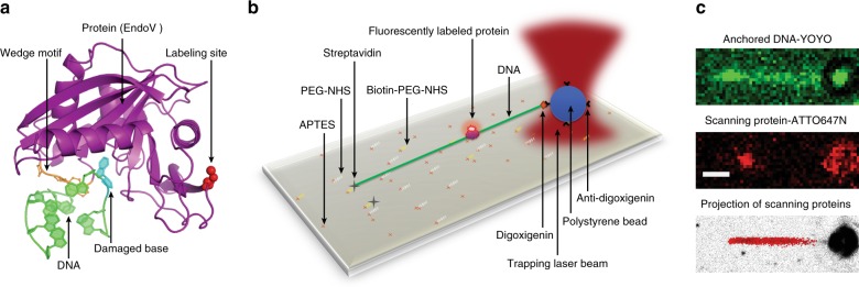
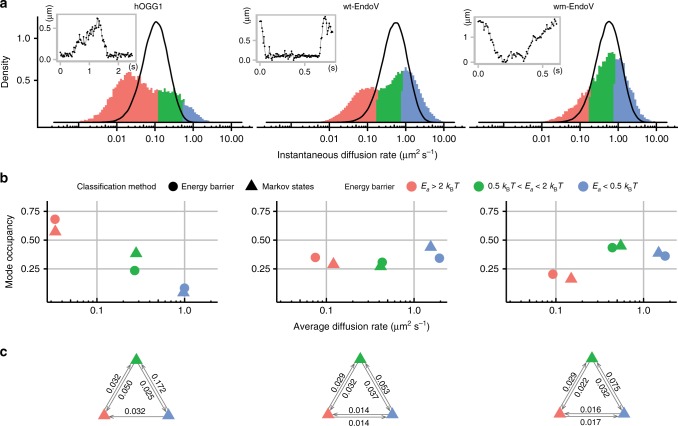
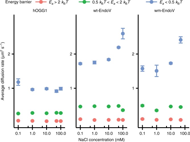
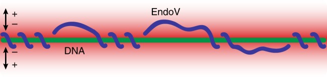
In order to preserve genomic stability, cells rely on various repair pathways for removing DNA damage. The mechanisms how enzymes scan DNA and recognize their target sites are incompletely understood. Here, by using high-localization precision microscopy along with 133 Hz high sampling rate, we have recorded EndoV and OGG1 interacting with 12-kbp elongated λ-DNA in an optical trap. EndoV switches between three distinct scanning modes, each with a clear range of activation energy barriers. These results concur with average diffusion rate and occupancy of states determined by a hidden Markov model, allowing us to infer that EndoV confinement occurs when the intercalating wedge motif is involved in rigorous probing of the DNA, while highly mobile EndoV may disengage from a strictly 1D helical diffusion mode and hop along the DNA. This makes EndoV the first example of a monomeric, single-conformation and single-binding-site protein demonstrating the ability to switch between three scanning modes.


Conflict of interest statement

The authors declare no competing interests. I.R. is currently employed by F. Hoffmann-La Roche (Roche Norge AS). The data included in this paper are based on research which has had no influence or involvement by F. Hoffmann-La Roche by any means. Any personal views of I.R. should not be understood or quoted as being made on behalf of or reflecting the position of F. Hoffmann-La Roche.

Figures

Fig. 1
Fig. 1
Protein–DNA interaction experiment. a Crystal structure of Thermotoga maritima (Tma) EndoV (purple) in complex with deaminated DNA (green). The protein labeling site (red spheres) does not interfere with DNA binding and damage recognition (cyan base in DNA). The PYIP-wedge motif (orange) consists of the four residues: Pro79, Tyr80, Ile81 and Pro82. b Single-molecule experimental setup. APTES (3-Aminopropyl)triethoxysilane, PEG-NHS N-hydroxylsuccinimide (NHS) functionalized polyethylene glycol, Biotin-PEG-NHS biotinylated PEG-NHS. The position of the polystyrene bead is controlled by a steerable optical trap, and used to linearize the DNA. c Upper panel: epifluorescence microscopy image of linearized DNA labeled with intercalating dye YOYO-1 (not present during the protein scanning experiments). Middle panel: a single fluorescently labeled EndoV molecule interacting with linearized DNA. Lower panel: projection of all detected trajectories of one data set of EndoV along the DNA. White scale bar equals 1 µm
Fig. 2
Fig. 2
Diffusion analysis. a Distribution of instantaneous diffusion coefficients computed for hOGG1 (697 trajectories) with sliding window of 117.5 ms, and for wt- and wm-EndoV (wt-EndoV: 3443 trajectories, and wm-EndoV: 1099 trajectories) using sliding window of 37.5 ms. Insets: examples of single trajectories for each protein with time on the x-axis and displacement on the y-axis. b Comparison of classifications based on activation energy barrier (circles) and on Markov hidden state models (triangles), showing strong agreement between methods. The state occupancy is the proportion of time that proteins spend in each of the classified modes. The standard error of the mean of the average diffusion rate for all points is below 0.02 µm2 s−1. c Inter-state transition probabilities of the hidden Markov models. Arrows between triangles show transition probabilities for the transitions between the respective scanning modes, colored according to the energy barrier
Fig. 3
Fig. 3
Diffusion rate analysis by buffer salt concentration. Average diffusion rates for each mode of diffusion as a function of buffer salt concentration. Error bars show the standard error of the mean (SEM) and are not visible when smaller than the size of the points in the plot
Fig. 4
Fig. 4
Model of interspersed helical sliding and hopping on DNA. The protein trajectory (blue trace) is confined within the radial electrostatic field (red to white) surrounding the DNA (green) to achieve rapid reassociation to DNA and efficient target localization during DNA scanning. Number of turns and length of hopping steps are unknown parameters and not to scale in this scheme

References

    1. Friedberg, E. C. et al. DNA Repair and Mutagenesis (ASM Press, Washington, 2006).
    1. Hoeijmakers JHJ. DNA damage, aging, and cancer. N. Engl. J. Med. 2009;361:1475–1485. doi: 10.1056/NEJMra0804615. - DOI - PubMed
    1. Wood RD, Mitchell M, Sgouros J, Lindahl T. Human DNA repair genes. Science. 2001;291:1284–1289. doi: 10.1126/science.1056154. - DOI - PubMed
    1. Berg OG, Winter RB, von Hippel PH. Diffusion-driven mechanisms of protein translocation on nucleic acids. 1. Models and theory. Biochemistry. 1981;20:6929–6948. doi: 10.1021/bi00527a028. - DOI - PubMed
    1. Riggs AD, Bourgeois S, Cohn M. The lac represser-operator interaction. III. Kinetic studies. J. Mol. Biol. 1970;53:401–417. doi: 10.1016/0022-2836(70)90074-4. - DOI - PubMed

Publication types

MeSH terms

Substances