Review
. 2019 Apr;38(16):2885-2898.
doi: 10.1038/s41388-018-0637-x. Epub 2018 Dec 19.

An evolving story of the metastatic voyage of ovarian cancer cells: cellular and molecular orchestration of the adipose-rich metastatic microenvironment

Affiliations
Review

An evolving story of the metastatic voyage of ovarian cancer cells: cellular and molecular orchestration of the adipose-rich metastatic microenvironment

Takeshi Motohara et al. Oncogene. 2019 Apr.

Erratum in

Abstract

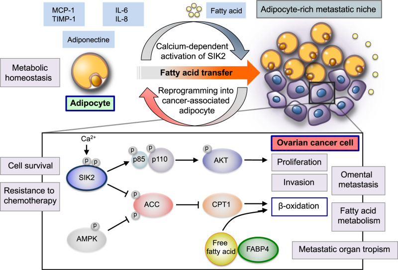
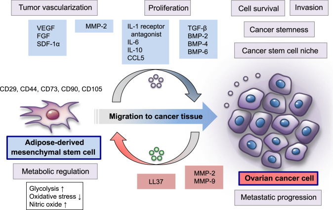
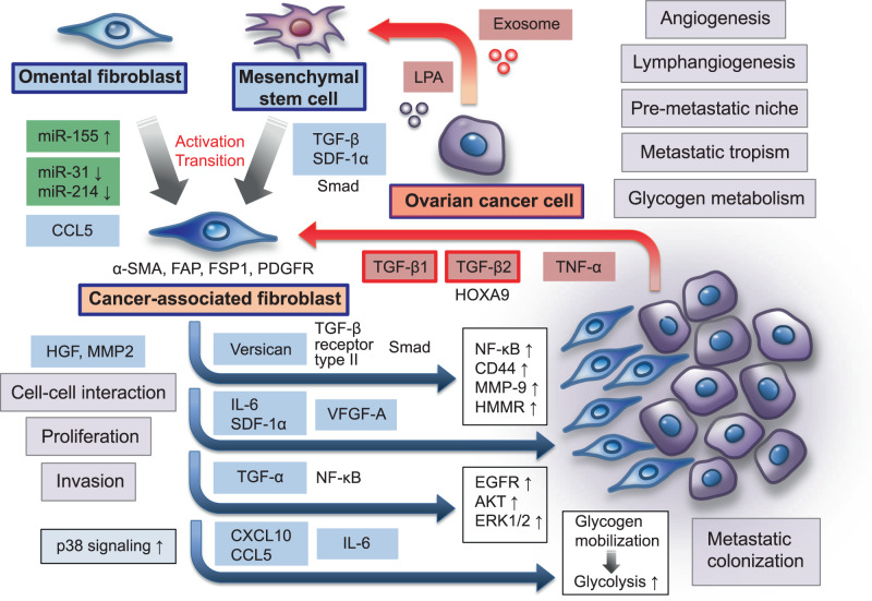
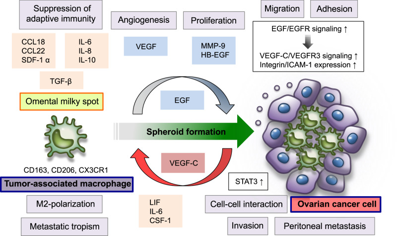
Metastasis is a complex multistep process that involves critical interactions between cancer cells and a variety of stromal components in the tumor microenvironment, which profoundly influence the different aspects of the metastatic cascade and organ tropism of disseminating cancer cells. Ovarian cancer is the most lethal gynecological malignancy and is characterized by peritoneal disseminated metastasis. Evidence has demonstrated that ovarian cancer possesses specific metastatic tropism for the adipose-rich omentum, which has a pivotal role in the creation of the metastatic tumor microenvironment in the intraperitoneal cavity. Considering the distinct biology of ovarian cancer metastasis, the elucidation of the cellular and molecular mechanisms underlying the reciprocal interplay between ovarian cancer cells and surrounding stromal cell types in the adipose-rich metastatic microenvironment will provide further insights into the development of novel therapeutic approaches for patients with advanced ovarian cancer. Herein, we review the biological mechanisms that regulate the highly orchestrated crosstalk between ovarian cancer cells and various cancer-associated stromal cells in the metastatic tumor microenvironment with regard to the omentum by illustrating how different stromal cells concertedly contribute to the development of ovarian cancer metastasis and metastatic tropism for the omentum.


Conflict of interest statement

The authors declare that they have no conflict of interest.

Figures

Fig. 1
Fig. 1
Adipocytes represent a central player in the creation of the metabolic tumor microenvironment in the omentum during ovarian cancer metastasis. Adipocytes are reprogrammed into cancer-associated adipocytes by cancer-derived mediators. These activated adipocytes release a wealth of lipids and various adipokines, including IL-6, IL-8, MCP-1, and TIMP-1, which contribute to the establishment of the omental metastatic niche for ovarian cancer. FABP4, which is an intracellular chaperone for free fatty acids, regulates lipolysis in adipocytes and β-oxidation in ovarian cancer cells, and plays a crucial role in the interaction of ovarian cancer cells with adipocytes, promoting omental metastasis. In addition, omental adipocytes induce the calcium-dependent activation and autophosphorylation of SIK2 in ovarian cancer cells and activate cancer cell proliferation through the PI3K/AKT pathway. SIK2 can increase ovarian cancer cell fatty acid oxidation by augmenting AMPK-induced ACC phosphorylation and activating CPT1 during peritoneal disseminated metastasis. Thus, the highly orchestrated crosstalk between ovarian cancer cells and omental adipocytes induces metabolic synergies by reprogramming fatty acid metabolism and tumor-promoting signaling pathways that enhance the proliferation, invasion, and metastatic progression of ovarian cancer cells with specific metastatic tropism for the omentum
Fig. 2
Fig. 2
MSCs are an important cellular component of the omental metastatic microenvironment of ovarian cancer. Omental adipose-derived MSCs promote tumor vascularization via the upregulation of VEGF, FGF, and SDF1-α, thereby increasing ovarian cancer cell survival in the metastatic tumor microenvironment. These MSCs can enhance glycolysis and suppress oxidative stress in ovarian cancer cells by regulating nitric oxide levels during the establishment of the omental metastatic niche. In addition, adipose-derived MSCs promote ovarian cancer cell proliferation and metastasis by elevating the expression of MMP2 and MMP-9. Ovarian cancer cells possess the ability to recruit cancer-associated MSCs to the metastatic tumor microenvironment by producing the pro-tumorigenic peptide LL-37. LL37 activates MSCs to secrete a variety of inflammatory and pro-angiogenic factors, including IL-1 receptor antagonist, IL-6, IL-10, CCL5, VEGF, and MMP-2, which are involved in metastatic progression of ovarian cancer cells. Ovarian cancer-associated MSCs, determined by CD44, CD73, and CD90 expressions, play a key role in the formation of the cancer stem cell niche microenvironment in the intraperitoneal cavity, and promote ovarian cancer growth, correlated with enhanced cancer stem cell properties. Mechanistically, these cancer-associated MSCs are involved in the upregulation of the TGF-β superfamily/ BMP family members, especially BMP2, BMP4, and BMP6, and thereby accelerate the aggressiveness of ovarian cancer cells
Fig. 3
Fig. 3
Fibroblasts play a crucial role in the establishment of the omental tumor microenvironment of ovarian cancer. Omental fibroblasts contribute to the creation of a pre-metastatic niche and influence tropism for the omentum and the metastatic colonization of ovarian cancer cells. Ovarian cancer-derived LPA and exosomes stimulate the differentiation of adipose-derived MSCs to CAFs, which are characterized by α-SMA, FAP, FSP1, and PDGFR expressions, by activating TGF-β-related signaling pathways. In addition, ovarian cancer cells can reprogram normal omental fibroblasts to CAFs via the upregulation of miR-155 and the downregulation of miR-31 and miR-214. This promotes tumor growth through increased secretion of CCL5. Ovarian cancer-derived TGF-β1, TGF-β2, and TNF-α, are involved in stimulating the production of various tumor-promoting factors, such as versican, IL-6, SDF-1α, and VEGF-A, in the metastatic tumor microenvironment. In particular, CAF-derived TGF-α promotes the metastatic colonization of ovarian cancer cells via the activation of EGFR, AKT, and ERK1/2 signaling pathways. Metastasizing ovarian cancer cells can activate p38 signaling in omental CAFs, and CAF-derived p38-regulated cytokines and chemokines, including IL-6, CXCL10, and CCL5, induce cancer cells to metabolize glycogen through glycolysis, which mediates energy production and accelerates the aggressiveness of ovarian cancer cells. Thus, in the intraperitoneal metastatic microenvironment, CAFs coevolve with ovarian cancer cells and govern the metastatic cascade, including the adhesion, proliferation, invasion, and colonization of metastatic sites
Fig. 4
Fig. 4
TAMs are involved in the establishment of the inflammatory and immunosuppressive tumor microenvironment during ovarian cancer peritoneal metastasis. Omental milky spots serve as the major sources of intraperitoneal macrophages and play a crucial role in the regulation of metastatic tropism for the omentum. Ovarian cancer cells polarize TAMs to the M2 phenotype, characterized by expressions of CD163, CD206, and CX3CR1, by upregulating LIF, IL-6, and CSF-1 in the specific tumor microenvironment of the intraperitoneal cavity; this microenvironment is modulated by a large number of soluble factors, such as IL-6, IL-8, IL-10, TGF-β, CCL18, CCL22, SDF-1α, VFGF, MMP-9, and HB-EGF. Intraperitoneal M2 macrophages can activate STAT3 signaling pathways in ovarian cancer cells. These pathways are associated with cell–cell interactions between cancer cells and M2 macrophages. Furthermore, these mutual interactions between ovarian cancer cells and macrophages are involved in formation of multicellular spheroids in the malignant ascites of ovarian cancer. EGF secreted by M2 macrophage activates EGFR on ovarian cancer cells, which in turn upregulates VEGF-C/VFGFR3 signaling pathways and enhances integrin/ICAM-1 expression in the intraperitoneal tumor microenvironment, promoting ovarian cancer proliferation, invasion, and intraperitoneal dissemination

References

    1. Bast RC, Jr., Hennessy B, Mills GB. The biology of ovarian cancer: new opportunities for translation. Nat Rev Cancer. 2009;9:415–28. doi: 10.1038/nrc2644. - DOI - PMC - PubMed
    1. Agarwal R, Kaye SB. Ovarian cancer: strategies for overcoming resistance to chemotherapy. Nat Rev Cancer. 2003;3:502–16. doi: 10.1038/nrc1123. - DOI - PubMed
    1. Patch AM, Christie EL, Etemadmoghadam D, Garsed DW, George J, Fereday S, et al. Whole-genome characterization of chemoresistant ovarian cancer. Nature. 2015;521:489–94. doi: 10.1038/nature14410. - DOI - PubMed
    1. Mills K, Fuh K. Recent advances in understanding, diagnosing, and treating ovarian cancer. F1000Res. 2017;6:84. doi: 10.12688/f1000research.9977.1. - DOI - PMC - PubMed
    1. Bowtell DD, Bohm S, Ahmed AA, Aspuria PJ, Bast RC, Jr., Beral V, et al. Rethinking ovarian cancer II: reducing mortality from high-grade serous ovarian cancer. Nat Rev Cancer. 2015;15:668–79. doi: 10.1038/nrc4019. - DOI - PMC - PubMed

Publication types