. 2019 Jan:99:1-26.
doi: 10.1016/j.pmatsci.2018.07.005. Epub 2018 Jul 11.

Nanomaterials as photothermal therapeutic agents

Affiliations

Nanomaterials as photothermal therapeutic agents

Junqi Chen et al. Prog Mater Sci. 2019 Jan.

Abstract

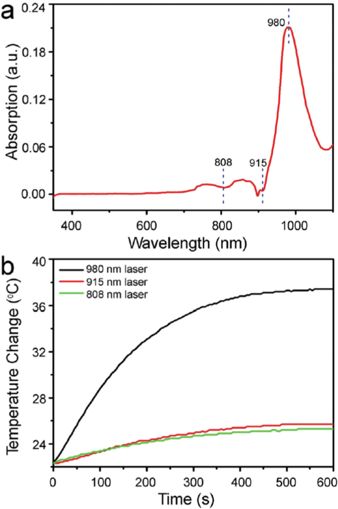
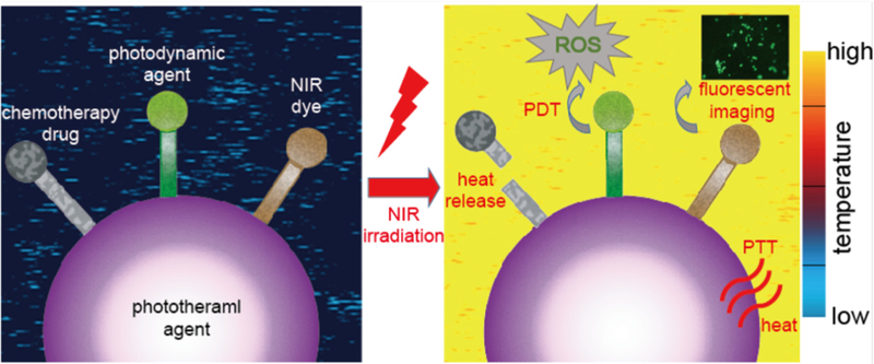
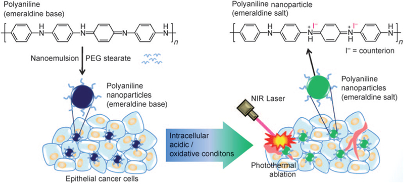
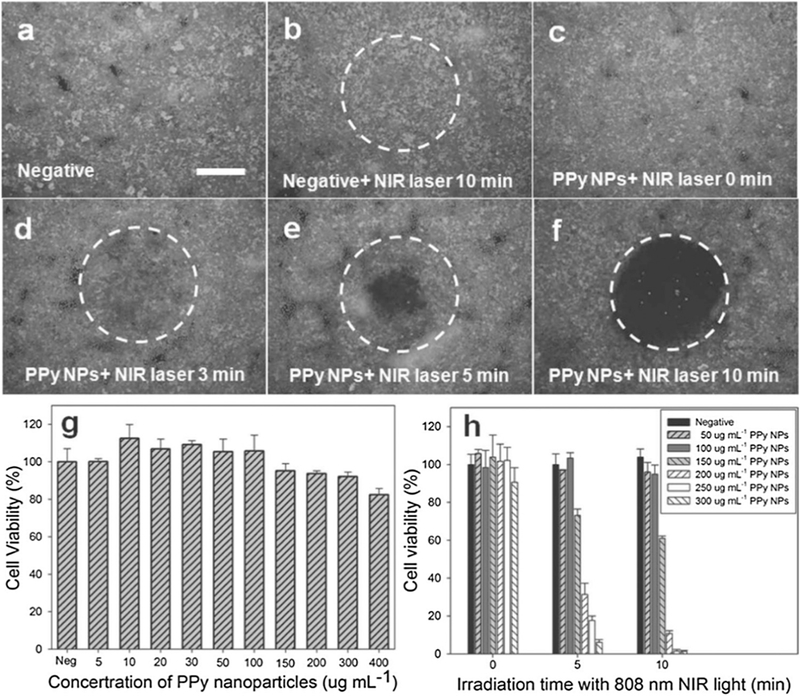
Curing cancer has been one of the greatest conundrums in the modern medical field. To reduce side-effects associated with the traditional cancer therapy such as radiotherapy and chemotherapy, photothermal therapy (PTT) has been recognized as one of the most promising treatments for cancer over recent years. PTT relies on ablation agents such as nanomaterials with a photothermal effect, for converting light into heat. In this way, elevated temperature could kill cancer cells while avoiding significant side effects on normal cells. This theory works because normal cells have a higher heat tolerance than cancer cells. Thus, nanomaterials with photothermal effects have attracted enormous attention due to their selectivity and non-invasive attributes. This review article summarizes the current status of employing nanomaterials with photothermal effects for anti-cancer treatment. Mechanisms of the photothermal effect and various factors affecting photothermal performance will be discussed. Efficient and selective PTT is believed to play an increasingly prominent role in cancer treatment. Moreover, merging PTT with other methods of cancer therapies is also discussed as a future trend.

Keywords: Cancer; Nanomaterials; Photothermal therapy.


Figures

Fig. 1.
Fig. 1.
(a) UV-vis absorption spectrum of the pure water. (b) Temperature elevation of the pure water under the irradiation of 808 nm, 915 nm and 980nm lasers with the same power density of 1.0Wcm−2 for 10min [73].
Fig. 2.
Fig. 2.
Schematic illustration of photothermal therapy and the synergic medical applications combined with phtotothermal therapy and other light- induced treatment, such as light-induced release of drugs for chemotherapy, photodynamic therapy and NIR fluorescent imaging. The color of background indicates the change of temperature during the photothermal therapy according to the color map on the right.
Fig. 3.
Fig. 3.
UV-vis absorption spectrum of gold nanorods with an average aspect ratio of 3.3. The band at 740 nm is referred to as the longitudinal plasmon absorption [97].
Fig. 4.
Fig. 4.
Representative scheme illustrating the fabrication of DNA-wrapped GNRs with DOX loading, and cellular mechanism for combinational chemo and PTA cancer therapy [104].
Fig. 5.
Fig. 5.
Schematic Illustration of the Stepwise Self-Assembly of CuS NPs to Primary Nanorods and Secondary Shuttle-like Bundles and the photothermal effect improvement from CuS NPs to Primary Nanorods and Secondary Shuttle-like Bundles [118].
Fig. 6.
Fig. 6.
UV-vis spectra of Cu2-xS NCs prepared by (A) sonoelectrochemical, (B) hydrothermal, and (C) thermolysis methods [119].
Fig. 7.
Fig. 7.
A proposed scheme to explain SWNT skin accumulation. The micro-vessels with diameters as small as 5 mm enriched in the skin dermis may tend to trap SWNTs overtime once nanotubes have an ultra-long blood circulation half-life [38].
Fig.8.
Fig.8.
The intracellular mechanisms of graphene-induced photothermal cell death. (A-D) U251 cells were exposed for 3 min to NIR laser (808 nm, 2Wcm−2) in the absence or presence of GPVP (10mg/ml). After 24h, flow cytometry was used to assess caspase activation (ApoStat staining; A), while the mitochondrial membrane potential (ΔΨ) (DePsipher staining; B), production of ROS (DHR staining; C) and superoxide (DHE staining; D) were measured after 4 h. The representative photomicrographs, histograms and dot plots are shown. The results are mean ± SD values from three experiments (*p < 0.05 refers to untreated cells and cells treated with NIR or GPVP alone; ANOVA). [152]
Fig. 9.
Fig. 9.
Schematic illustration of the preparation of organic photothermal agents based on polyaniline nanoparticles and their application for the photothermal ablation of epithelial cancer cells by NIR laser irradiation [164].
Fig. 10.
Fig. 10.
Photothermal destruction of Hela cells with or without PPy NPs and NIR laser (808 nm, 6Wx m−2) treatments (a, b, c, d, e and f). White circle indicates the laser spot; live/dead stain for viability. Scale bar = 800 μm. (g) Cell viability of HUVECs with 24 h exposure to various concentrations of PPy NPs. (h) Cell viability after treatment with different concentrations of PPy NPs and different NIR laser irradiation time [86].
Fig. 11.
Fig. 11.
Schematic illustration of photothermal effect of PPy-coated chainlike gold NPs [169].
Fig. 12.
Fig. 12.
Schematic illustration of the formation of TaOx@PPy NPs for combined CT/PA imaging and PTT [196].
Fig. 13.
Fig. 13.
(A) SERS spectra of the A549 cells with (a) and without (b) binding of the Rh6G-labeled aptamer-Ag-Au nanostructures. (c-h) SERS spectra of the NCIH157, NCI-H520, NCI-H1299, NCI-H446, MCF-7, and HeLa cells, respectively, after incubating with the Rh6G-labeled aptamer-Ag-Au nanostructures. (B) SERS spectra of the A549 cells after binding of the Rh6G-labeled aptamer-Ag-Au nanostructures at the cell number (cells per mL) of (a) ~ 1* 101, (b) 1 * 102, (c) 1 *103, (d) 1 * 104, (e) 1 * 105, and (f) 1* 106, respectively. (C) Dependence of the SERS signals at 1363 cm−1 on the A549 cell numbers (in cells per mL) [200].
Fig. 14.
Fig. 14.
NIR SERS imaging of 4T1 cells using GNR-PANI (a-c) or GNR-PPy (d-f) as nanotags. (g and h) SERS spectral lines acquired from the 4T1 cells incubated with GNR-PANI (c) and GNR-PPy (f), respectively. Black lines are the background lines. Scale bar: 10 μm [201].
Fig. 15.
Fig. 15.
Illustration of hyperthermia-driven transport. Mild heat is generated by near infrared laser irradiation aided by heat-absorbing gold nanorods (A). Effect on tumor transport was studied by intravital microscopy with images demonstrating increased macromolecular accumulation (fluorescent intensity) in tumors receiving heat treatment versus control (B). Mathematic modeling was further used to quantify enhancements which revealed that the rate of extravasation increased by 30% while the total macromolecule accumulation increased by 200% (C) [210].
Fig. 16.
Fig. 16.
(A) SEM images of freshly prepared core-shell plasmonic nanopopcorn. (B) EDX mapping shows the presence of Fe and Au in a core-shell nanoparticle. (C) Absorption spectra of core-shell nanoparticle, methylene blue, and methylene blue conjugated nanoplatform [221].
Fig. 17.
Fig. 17.
Schematic illustration of PS-loaded micelles integrating cyanine dye for dual-modal cancer imaging and synergistic therapy of PTT and PDT via an enhanced cytoplasmic delivery of PS [222].
Fig. 18.
Fig. 18.
Schematic illustration of synthesis and microstructure of reduced Graphene Oxide (rGO), Porous Silica nanoSheet (PSS), rGO@Silica nanoSheet (rGO@SS), GO@Porous Silica nanoSheet and nanocookie [228].
Fig. 19.
Fig. 19.
Schematic representation of five-function Fe3O4@Au nanorose for cancer cell targeting, MRI, optical imaging, photothermal and chemotherapy. Sgc8 aptamers are conjugated on the surface of nanoroses for targeting of CCRF-CEM cancer cells. Anticancer drug (Dox) is specifically delivered into cancer cells for chemotherapy and optical imaging. Fe3O4@Au nanorose is an efficient MRI agent and photothermal agent; therefore, MRI and photothermal therapy can be achieved for cancer cell imaging and therapy [230].
Fig. 20.
Fig. 20.
Schematic diagram of photosensitizer conjugation onto biological nanowire and its targeted photodynamic therapy [260].

Similar articles

Cited by

References

    1. Chen Y, Jungsuwadee P, Vore M, Butterfield D, St-Clair D. Collateral damage in cancer chemotherapy: oxidative stress in nontargeted tissues. Mol Interventions 2007;7:2653–63. - PubMed
    1. Edison M, Johns C. Acute and chronic cutaneous reactions to radiotherapy In: Cognetta AB, Mendenhall WM, editors. Radiation therapy for skin cancer. New York: Springer; 2013. p. 55–69.
    1. Matteini P, Tatini F, Cavigli L, Ottaviano S, Ghini G, Pini R. Graphene as a photothermal switch for controlled drug release. Nanoscale 2014;6:7947–53. - PubMed
    1. Tang S, Chen M, Zheng N. Sub-10-nm Pd nanosheets with renal clearance for efficient near-infrared photothermal cancer therapy. Small 2014;10:3139–44. - PubMed
    1. Huang X, Jain PK, El-Sayed IH, El-Sayed MA. Plasmonic photothermal therapy (PPTT) using gold nanoparticles. Lasers Med Sci 2008;23:217–28. - PubMed

LinkOut - more resources