. 2018 Nov 28:10:783-795.
doi: 10.2147/IJWH.S174734. eCollection 2018.

The umbilical cord, preeclampsia and the VEGF family

Affiliations

The umbilical cord, preeclampsia and the VEGF family

Mercedes Olaya-C et al. Int J Womens Health. .

Abstract

Introduction: The VEGF family has been identified as abnormal in preeclampsia (PE). Hypertensive disorders of pregnancy (HDP) are major contributors to maternal and neonatal morbidity and mortality worldwide; likewise, umbilical cord anatomical abnormalities (UCAA) are linked to poor neonatal outcomes. Based on the relationship described between PE and UCAA and the role of the VEGF family in PE, this study explored VEGF expression in placental and UC tissued from patients with PE and with UCAA.

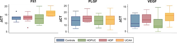
Methods: We performed an observational, analytical study on placentas, comparing protein and mRNA expression in four groups: patients with PE, patients with UC abnormalities, patients with both, and patients with none of them. Using immunohistochemistry, we studied VEGF A, VEGF R1 (FLT1), MMP1, and PLGF. With quantitative reverse transcription polymerase chain reaction we described mRNA expression of PLGF, VEGF and sFLT1, and sFLT1/PLGF ratio.

Results: Forty newborns were included. Sixty-seven percent of mothers and 45% of newborns developed no complications. Immunohistochemistry was performed on UC and placental disc paraffin-embedded tissue; in the latter, the mRNA of the VEGF family was also measured. Statistically significant differences were observed among different expressions in both HDP and UCAA groups. Interestingly, the UCAA group exhibited lower levels of sFLT1 and VEGF-A in comparison with other groups, with significant P-value for sFLT1 (P=0000.1).

Conclusion: The origin of UCAA abnormalities and their relation with HDP are still unknown. VEGF family alterations could be involved in both. This study provides the first approach related to molecules linked to UCAA.

Keywords: PLGF; VEGF; preeclampsia; sFLT1; stillbirth; umbilical cord.


Conflict of interest statement

Disclosure None of the authors has any potential or actual interests relevant to the topics discussed in this manuscript. The authors report no conflicts of interest in this work.

Figures

Figure 1
Figure 1
VEGF-A. Representative immunohistochemistry staining for VEGF-A in the placental disc. Positive expression demonstrated for smooth muscle of chorionic vessels, 10× (A), stromal chorionic cells, 20× (orange arrows), with positive internal control in chorionic endothelium (white arrow), and villous endothelial positivity (B), 20× (orange arrow), with negative internal control in syncytiotrophoblast (blue arrow) (C). Amnion negativity is shown 20× (D).
Figure 2
Figure 2
VEGF-A. Representative immunohistochemistry staining for VEGF-A in the umbilical cord. Note: Positive expression demonstrated for smooth muscle of artery 4× (A), smooth muscle of artery (blue arrows) and endothelium 10× (orange arrow) (B), amnion (orange arrows) 10× and stromal cells (blue arrows) 20× (C and D).
Figure 3
Figure 3
VEGF-R1. Representative immunohistochemistry staining for VEGF-R1 in the placental disc. Notes: Positive diffuse and strong expression demonstrated in syncytiotrophoblast 20× (B), extravillous trophoblast 4× (C), and decidua 20× (D). Chorionic plate is highlighted in (A) 10×, where trophoblast (orange arrows) and amnion (blue arrow) are prominent. Negative internal controls are seen in A and D (blue cells).
Figure 4
Figure 4
PLGF. Representative immunohistochemistry staining for PLGF. Notes: (A) Diffuse and strong positivity is seen in extravillous trophoblast, basal trophoblast, and syncytiotrophoblast. In (B) strong expression of this kind of trophoblast contrasts with weak expression in decidua. In C and D, umbilical cord shows strong and diffuse expression in the endothelium and the amniotic epithelium, even though it is present in smooth muscle cells, endothelium, and stromal cells.
Figure 5
Figure 5
MMP1. Representative immunohistochemistry staining for MMP1 in the placental disc. Notes: Positive expression demonstrated in trophoblast behind the basal plate 20× (A) (arrows) and smooth muscle of chorionic vessels 20× (B). Negative internal controls are shown (blue cells).
Figure 6
Figure 6
Immunohistochemistry summary. Notes: Galbraith’s graphic shows the compilation of the most representative results. LCL corresponds to minor CI and UCL corresponds to major CI. Abbreviations: HDP, hypertensive disorders of pregnancy; UCAA, umbilical cord anatomical alterations.
Figure 7
Figure 7
mRNA expression summary. Notes: Bar graphs (A) and box and whisker plots (B) show differences among groups: HDP, UCAA, HDPUC, and normal gestations (CTRL). The UCAA group was unexpectedly different; sFLT1 and VEGF-A expression was consistently lower than other groups. Abbreviations: CTRL, control; HDP, hypertensive disorders of pregnancy; HDPUC, HDP plus UCAA; UC, umbilical cord; UCAA, UC anatomical abnormalities.
Figure 8
Figure 8
mRNA expression summary. Notes: mRNA expression summary. Kruskal-Wallis test revealed significant differences upon comparing UCAA group in sFlt1 expression. In the y-axis, ΔCT. Abbreviations: HDP, hypertensive disorders of pregnancy; HDPUC, HDP plus UCAA; UC, umbilical cord; UCAA, umbilical cord anatomical abnormalities.

References

    1. US Preventive Services Task Force. Bibbins-Domingo K, Grossman DC, et al. Screening for preeclampsia: US preventive services task force recommendation statement. JAMA. 2017;317(16):1661–1667. - PubMed
    1. Bilano VL, Ota E, Ganchimeg T, Mori R, Souza JP. Risk factors of pre-eclampsia/eclampsia and its adverse outcomes in low- and middle-income countries: a WHO secondary analysis. PLoS One. 2014;9(3):e91198. - PMC - PubMed
    1. Foo L, Tay J, Lees CC, McEniery CM, Wilkinson IB. Hypertension in pregnancy: natural history and treatment options. Curr Hypertens Rep. 2015;17(5):36. - PubMed
    1. Shennan AH, Green M, Chappell LC. Maternal deaths in the UK: pre-eclampsia deaths are avoidable. Lancet. 2017;389(10069):582–584. - PubMed
    1. Steegers EA, von Dadelszen P, Duvekot JJ, Pijnenborg R. Pre-eclampsia. Lancet. 2010;376(9741):631–644. - PubMed

LinkOut - more resources