Review
. 2018 Dec 5:9:2802.
doi: 10.3389/fimmu.2018.02802. eCollection 2018.

Autoimmune Glial Fibrillary Acidic Protein Astrocytopathy: A Review of the Literature

Affiliations
Review

Autoimmune Glial Fibrillary Acidic Protein Astrocytopathy: A Review of the Literature

Fulan Shan et al. Front Immunol. .

Abstract

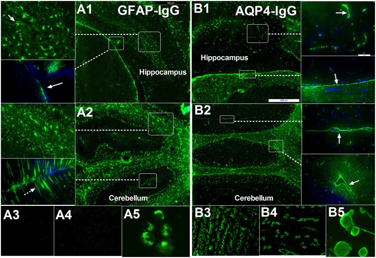
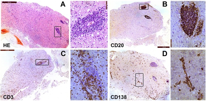
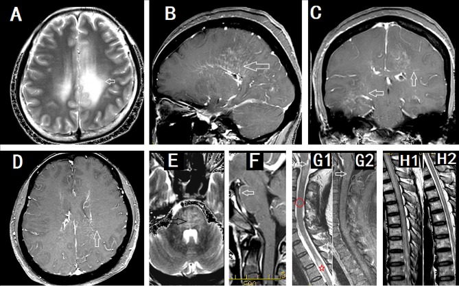
Autoimmune glial fibrillary acidic protein (GFAP) astrocytopathy is an autoimmune disease of the nervous system first defined in 2016. GFAP autoantibody, especially IgG that binds to GFAPα, has been reported in the cerebrospinal fluid (CSF) and serum of patients with GFAP astrocytopathy. The positive predictive value of GFAP antibody in the CSF is higher than in the serum. Tissue-based assay (TBA) and cell-based assay (CBA) are both recommended methods for the detection of GFAP antibody. GFAP astrocytopathy is accompanied by neoplasms, but the relationship between virus infection and GFAP astrocytopathy is unclear. GFAP antibody itself does not induce pathological changes; it is only a biomarker for the process of immune inflammation. The pathology of GFAP astrocytopathy in humans is heterogeneous. GFAP astrocytopathy is commonly diagnosed in individuals over 40 years old and most patients have an acute or subacute onset. Clinical manifestations include fever, headache, encephalopathy, involuntary movement, myelitis, and abnormal vision. Lesions involve the subcortical white matter, basal ganglia, hypothalamus, brainstem, cerebellum, and spinal cord. The characteristic MRI feature is brain linear perivascular radial gadolinium enhancement in the white matter perpendicular to the ventricle. Currently, there are no uniform diagnostic criteria or consensus for GFAP astrocytopathy and coexisting neural autoantibodies detected in the same patient make the diagnosis difficult. A standard treatment regimen is yet to be developed. Most GFAP astrocytopathy patients respond well to steroid therapy although some patients are prone to relapse or even die.

Keywords: antibody; astrocyte; astrocytopathy; glial fibrillary acidic protein; meningoencephalitis.


Figures

Figure 1
Figure 1
Comparison of immunofluorescence-pattern between GFAP-IgG and AQP4-IgG. (A1–A5) IgG from patient with GFAP astrocytopathy. (A1) The IgG is bound to the foot process at pia (arrow) and astrocyte body in hippocampus. (A2) IF pattern in different layer of cerebellum: (1) The IgG is bound to the astrocyte body in different layer, especially white matter (arrow); (2) it was detected in molecular layers with bergmann radial pattern (arrow). (A3,A4) no pattern-specific staining was detected in the kidney and stomach tissue but positive for GFAP-transinfected HEK-293 cell. (B1–B5) IgG from a positive AQP4-IgG NMO patient. (B1) The IgG is bound to the cell with the foot process around the microvessels (arrow) in brain and pia (arrow). (B2) Anti-AQP4 pattern located at the border between two molecular layers with Virchow-Robin space profile (arrow) without bergmann radial pattern. (B3–B5) Pattern-specific staining was detected in the stomach, kidney tissue, and AQP4-transinfected HEK-293 cell.
Figure 3
Figure 3
Neuropathological features of a patient with encephalitis. The case involved a woman, whose age at onset was 69 years. In January 2015, she experienced psychosis, dyskinesia, persistent fever, and paralysis of a right-side limb. Antibody detection and biopsy were conducted before the IVMP and IVIG treatment. She then received treatment with methylprednisolone and azathioprine. She did not response well to this treatment and died of acute respiratory failure 1 year later. (A) Hematoxylin-eosin staining of brain biopsy tissue shows extensive infiltration of inflammatory cells; (B) Immunostaining for anti-CD20 is mainly around the vessels in lesions; (C) Immunostaining for anti-CD3 is sparse throughout the lesion, with some staining around the large vessel; (D) CD138+ cells are present in perivascular, Virchow–Robin, and interstitial spaces.
Figure 2
Figure 2
Imaging findings in patients with GFAP astrocytopathy. (A–D) were from a female meningoencephalitis patient. (A) MR images showing extensive abnormalities in the white matter around the ventricle (arrow). (B) Sagittal section showed linear perivascular radial gadolinium enhancement in the white matter perpendicular to the ventricle(arrow). (C,D) Coronal section (C) and cross section (D) showed vessel-like enhancement (arrows). (E) and (F) from a male meningoencephalitis patient showed pons abnormality (black arrow) and pia enhancement (white arrow). (G1) and (G2) were from a female with myelitis. (G1) Cervical lesion extended to the area postrema of medulla(arrow), sparse cervical abnormality(red round area) and thoracic LESCLs (star marker). (G2) Slightly enhancement in medulla (arrow). (H1) and (H2) were from a male meningoencephalomyelitis patient. Longitudinal extensive lesions in the whole spinal cord (H1) and soon recovery after the treatment (H2).

References

    1. Lucchinetti CF, Guo Y, Popescu BF, Fujihara K, Itoyama Y, Misu T. The pathology of an autoimmune astrocytopathy: lessons learned from neuromyelitis optica. Brain Pathol. (2014) 24:83–97. 10.1111/bpa.12099 - DOI - PMC - PubMed
    1. Fang B, McKeon A, Hinson SR, Kryzer TJ, Pittock SJ, Aksamit AJ, et al. Autoimmune glial fibrillary acidic protein astrocytopathy: a novel meningoencephalomyelitis. JAMA Neurol. (2016) 73:1297–307. 10.1001/jamaneurol.2016.2549 - DOI - PubMed
    1. Flanagan EP, Hinson SR, Lennon VA, Fang B, Aksamit AJ, Morris PP, et al. Glial fibrillary acidic protein immunoglobulin G as biomarker of autoimmune astrocytopathy: analysis of 102 patients. Ann Neurol. (2017) 81:298–309. 10.1002/ana.24881 - DOI - PubMed
    1. Yang X, Liang J, Huang Q, Xu H, Gao C, Long Y, et al. Treatment of autoimmune glial fibrillary acidic protein astrocytopathy: follow-up in 7 cases. Neuroimmunomodulation (2017) 24:113–9. 10.1159/000479948 - DOI - PubMed
    1. Long Y, Liang J, Xu H, Huang Q, Yang J, Gao C, et al. Autoimmune glial fibrillary acidic protein astrocytopathy in Chinese patients: a retrospective study. Eur J Neurol. (2018) 25:477–83. 10.1111/ene.13531 - DOI - PubMed

Publication types

MeSH terms