

Femoroacetabular impingement as a complication of acetabular fracture fixation
- PMID: 30569011
- PMCID: PMC6286667
- DOI: 10.1016/j.tcr.2018.07.003
Femoroacetabular impingement as a complication of acetabular fracture fixation
Abstract
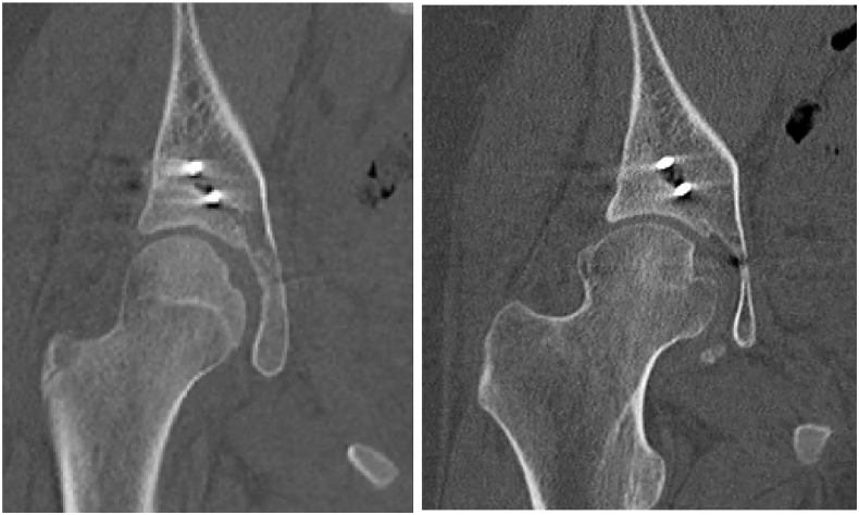
Case: We present the case of a thirteen-year-old female who sustained a posterior wall acetabular fracture dislocation. She underwent urgent closed reduction and subsequent uncomplicated open reduction and internal fixation. Post reduction computed tomography demonstrated a concentrically reduced hip joint with no evidence of femoroacetabular impingement (FAI). She subsequently healed her fracture and returned to running activities; however, one year later presented with aching pain in her thigh. Radiographs demonstrated the development of a large osseous prominence on her anterolateral femoral neck consistent with femoroacetabular impingement. Based on these findings she was evaluated by a hip preservation specialist. She subsequently underwent successful hip arthroscopy for labral repair and femoral osteochondroplasty. She was eventually able to return to running sports with little pain.
Summary: We present a case of FAI presenting as a complication of acetabular fracture fixation. This should be discussed with patients presenting with traumatic hip dislocations as a possible complication of surgical fixation or possibly of the injury itself.
Keywords: Acetabular fracture; Complication; Femoroacetabular impingement; Hip arthroscopy.
Figures


References
-
- Philippon M.J. Arthroscopic findings following traumatic hip dislocation in 14 professional athletes. Arthroscopy. 2009;25(2):169–174. - PubMed
-
- Philippon M.J. Arthroscopic management of femoroacetabular impingement: osteoplasty technique and literature review. Am. J. Sports Med. 2007;35(9):1571–1580. - PubMed
-
- Tonnis D., Heinecke A. Acetabular and femoral anteversion: relationship with osteoarthritis of the hip. J. Bone Joint Surg. Am. 1999;81(12):1747–1770. - PubMed
-
- Yanke A.B. Sex differences in patients with CAM deformities with femoroacetabular impingement: 3-dimensional computed tomographic quantification. Arthroscopy. 2015;31(12):2301–2306. - PubMed
-
- Matta J.M. Fractures of the acetabulum: accuracy of reduction and clinical results in patients managed operatively within three weeks after the injury. J. Bone Joint Surg. Am. 1996;78(11):1632–1645. - PubMed
Publication types
LinkOut - more resources
Full Text Sources
