. 2019 Feb;43(2):749-760.
doi: 10.3892/ijmm.2018.4034. Epub 2018 Dec 18.

DDAH2 alleviates myocardial fibrosis in diabetic cardiomyopathy through activation of the DDAH/ADMA/NOS/NO pathway in rats

Affiliations

DDAH2 alleviates myocardial fibrosis in diabetic cardiomyopathy through activation of the DDAH/ADMA/NOS/NO pathway in rats

Zhen-Dong Zhu et al. Int J Mol Med. 2019 Feb.

Abstract

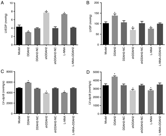
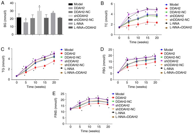
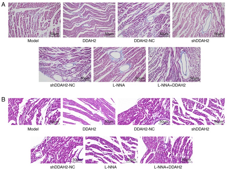
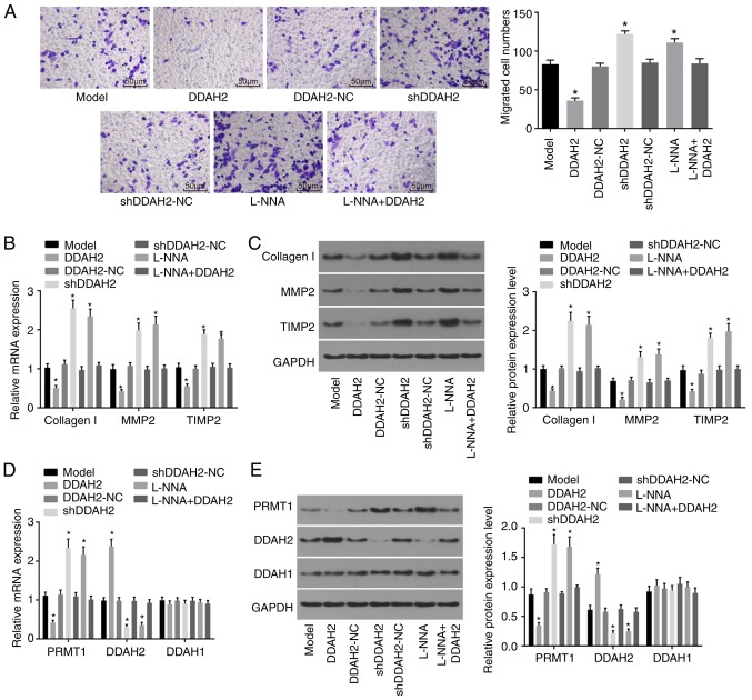
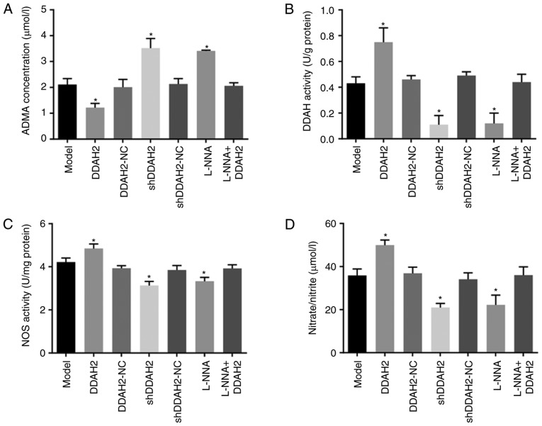
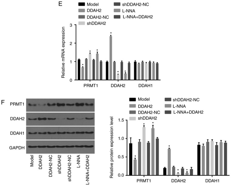
Diabetic cardiomyopathy (DCM) is a form of idiopathic heart disease, with signs including hypertrophy of myocardial cells, hypertension‑independent fibrosis and coronary artery disease. Considering the involvement of dimethylarginine dimethylaminohydrolase 2 (DDAH2) in diabetes, it was hypothesized that DDAH2 may be beneficial to cardiac function and myocardial fibrosis during the progression of DCM with involvement of the DDAH/asymmetric NG, NGdimethyl‑L‑arginine (ADMA)/nitric oxide synthase (NOS)/nitric oxide (NO) signaling pathway. Following establishment of diabetic rat models, diabetes‑related blood biochemical indices and cardiac function were measured in diabetic rats treated with lentivirus expressing DDAH2, short hairpin RNA against DDAH2, or L‑NNA (inhibitor of NOS) to identify the roles of DDAH2 in DCM. The functional roles of DDAH2 in DCM were further determined through detection of the levels of collagen I, matrix metalloproteinase 2 (MMP2) and tissue inhibitor of metalloproteinase 2 (TIMP2). The H9C2 myocardial cell line was selected for in vitro experiments. The effects of DDAH2 on the migration of myocardial cells under high glucose conditions were also examined. To further investigate the underlying regulatory mechanism of DDAH2 in DCM, the contents of ADMA and NO, and the activities of DDAH and NOS were observed. The DCM model rats treated with DDAH2 exhibited reduced left ventricular end‑diastolic pressure, and decreased blood glucose, total cholesterol, triglyceride, fasting blood glucose, and fasting insulin levels, but exhibited increased left ventricular systolic pressure and maximum rate of left ventricular pressure rise/fall levels in myocardial tissues. Myocardial cells under high glucose conditions treated with DDAH2 showed reductions in collagen I, MMP2 and TIMP2, indicating that DDAH2 reduced cell migration. Decreased levels of ADMA and NO but increased levels of DDAH and NOS were observed following treatment with DDAH2, indicating that the DDAH/ADMA/NOS/NO pathway was activated. These results reveal that the overexpression of DDAH2 attenuates myocardial fibrosis and protects against DCM through activation of the DDAH/ADMA/NOS/NO pathway in DCM rats. These results indicate that DDAH2 is a potential therapeutic candidate for the treatment of DCM.


Figures

Figure 1
Figure 1
DDAH2 exerts a protective effect on cardiac function in diabetic rats with myocardial fibrosis. (A) LVEDP level in diabetic rats following 12 weeks of treatment with lentivirus expressing DDAH2 or shDDAH2, and/or L-NNA. (B) LVSP level in diabetic rats following 12 weeks of treatment with lentivirus expressing DDAH2 or shDDAH2, and/or L-NNA. (C) LV + dp/dt level in diabetic rats following 12 weeks of treatment with lentivirus expressing DDAH2 or shDDAH2, and/or L-NNA. (D) LV-dp/dt level in diabetic rats following 12 weeks of treatment with lentivirus expressing DDAH2 or shDDAH2, and/or L-NNA. Data are presented as the mean ± standard deviation and analyzed by the one-way analysis of variance. All data are representative of three independent experiments. n=5. *P<0.05, vs. model group. DDAH2, dimethylarginine dimethylaminohydrolase 2; NC, negative control; sh, short hairpin RNA; LVEDP, left ventricular end-diastolic pressure, LVSP, left ventricular systolic pressure, LV ± dp/dt, maximum rate of left ventricular pressure rise/fall.
Figure 2
Figure 2
DDAH2 positively regulates blood biochemical indicators in diabetic rats with myocardial fibrosis. (A) BG level in diabetic rats following 5-20 weeks of treatment with lentivirus expressing DDAH2 or shDDAH2, and/or L-NNA. (B) TC level in diabetic rats following 5-20 weeks of treatment with lentivirus expressing DDAH2 or shDDAH2, and/or L-NNA. (C) TG level in diabetic rats following 5-20 weeks of treatment with lentivirus expressing DDAH2 or shDDAH2, and/or L-NNA. (D) FBG level in diabetic rats following 5-20 weeks of treatment with lentivirus expressing DDAH2 or shDDAH2, and/or L-NNA. (E) FINS in diabetic rats following 5-20 weeks of treatment with lentivirus expressing DDAH2 or shDDAH2, and/or L-NNA. *P<0.05. vs. model group. Data are presented as the mean ± standard deviation; data at the same time point in different groups were analyzed by one-way ANOVA; data at different time points were analyzed by repeated measure ANOVA. All data are representative of three independent experiments. n=5. DDAH2, dimethylarginine dimethylaminohydrolase 2; NC, negative control; sh, sort hairpin RNA; BG, blood glucose; TC, total cholesterol; TG, triglyceride; FBG, fasting blood glucose; FINS, fasting insulin; ANOVA, analysis of variance.
Figure 3
Figure 3
DDAH2 improves myocardial fibrosis in diabetic rats. (A) Masson staining results of diabetic rats following 20 weeks of treatment with lentivirus expressing DDAH2 or shDDAH2, and/or L-NNA in each group (magnification, ×400). (B) hematoxylin and eosin staining results of diabetic rats following 20 weeks of treatment with lentivirus expressing DDAH2 or shDDAH2, and/or L-NNA in each group (magnification, ×200). DDAH2, dimethylarginine dimethylaminohydrolase 2; NC, negative control; sh, short hairpin RNA.
Figure 4
Figure 4
DDAH2 inhibits myocardial cell migration. (A) Migration ability of H9C2 myocardial cells under high glucose conditions following 48 h of treatment with lentivirus expressing DDAH2 or shDDAH2, and/or L-NNA (magnification, ×200). (B) mRNA levels of collagen I, MMP2 and TIMP in H9C2 myocardial cells under high glucose conditions following 48 h of treatment with lentivirus expressing DDAH2 or shDDAH2, and/or L-NNA. (C) Protein levels of collagen I, MMP2 and TIMP in H9C2 myocardial cells under high glucose conditions following 48 h of treatment with lentivirus expressing DDAH2 or shDDAH2, and/or L-NNA. (D) mRNA expression of PRMT1, DDAH2 and DDAH1 in H9C2 myocardial cells under high glucose condition following 48 h of treatment with lentivirus expressing DDAH2 or shDDAH2, and/or L-NNA (E) Protein expression of PRMT1, DDAH2 and DDAH1 in H9C2 myocardial cells under high glucose conditions following 48 h of treatment with lentivirus expressing DDAH2 or shDDAH2, and/or L-NNA. Data are presented as the mean ± standard deviation and analyzed by one-way analysis of variance. All data are representative of three independent experiments. *P<0.05, vs. model group. DDAH, dimethylarginine dimethylaminohydrolase; PRMT1, protein arginine N-methyltransferase 1; NC, negative control; sh, short hairpin RNA; MMP2, matrix metalloproteinase 2; TIMP2, tissue inhibitor of metalloproteinase 2; GAPDH, glyceraldehyde-3-phosphate dehydrogenase.
Figure 5
Figure 5
DDAH2 activates the DDAH/ADMA/NOS/NO pathway in diabetic rats with myocardial fibrosis. (A) Serum ADMA content in diabetic rats following 20 weeks of treatment with lentivirus expressing DDAH2 or shDDAH2, and/or L-NNA. (B) DDAH activity in myocardial tissues of diabetic rats following 20 weeks of treatment with lentivirus expressing DDAH2 or shDDAH2, and/or L-NNA. (C) NOS activity in myocardial tissues of diabetic rats following 20 weeks of treatment with lentivirus expressing DDAH2 or shDDAH2, and/or L-NNA. (D) NO content in myocardial tissues of diabetic rats following 20 weeks of treatment with lentivirus expressing DDAH2 or shDDAH2, and/or L-NNA. (E) mRNA and (F) protein levels of PRMT1, DDAH2 and DDAH1 in myocardial tissues of rats following 20 weeks of treatment with lentivirus expressing DDAH2 or shDDAH2, and/or L-NNA. Data are presented as the mean ± standard deviation and were analyzed by one-way analysis of variance. *P<0.05, vs. model group All data are representative of three independent experiments. DDAH, dimethylarginine dimethylaminohydrolase; ADMA, asymmetric NG, NGdimethyl-L-arginine; PRMT1, protein arginine N-methyltransferase 1; NC, negative control; shRNA, short hairpin RNA; ADMA, asymmetric dimethylarginine; NOS, nitric oxide synthase; NO, nitric oxide; GAPDH, glyceraldehyde-3-phosphate dehydrogenase.
Figure 5
Figure 5
DDAH2 activates the DDAH/ADMA/NOS/NO pathway in diabetic rats with myocardial fibrosis. (A) Serum ADMA content in diabetic rats following 20 weeks of treatment with lentivirus expressing DDAH2 or shDDAH2, and/or L-NNA. (B) DDAH activity in myocardial tissues of diabetic rats following 20 weeks of treatment with lentivirus expressing DDAH2 or shDDAH2, and/or L-NNA. (C) NOS activity in myocardial tissues of diabetic rats following 20 weeks of treatment with lentivirus expressing DDAH2 or shDDAH2, and/or L-NNA. (D) NO content in myocardial tissues of diabetic rats following 20 weeks of treatment with lentivirus expressing DDAH2 or shDDAH2, and/or L-NNA. (E) mRNA and (F) protein levels of PRMT1, DDAH2 and DDAH1 in myocardial tissues of rats following 20 weeks of treatment with lentivirus expressing DDAH2 or shDDAH2, and/or L-NNA. Data are presented as the mean ± standard deviation and were analyzed by one-way analysis of variance. *P<0.05, vs. model group All data are representative of three independent experiments. DDAH, dimethylarginine dimethylaminohydrolase; ADMA, asymmetric NG, NGdimethyl-L-arginine; PRMT1, protein arginine N-methyltransferase 1; NC, negative control; shRNA, short hairpin RNA; ADMA, asymmetric dimethylarginine; NOS, nitric oxide synthase; NO, nitric oxide; GAPDH, glyceraldehyde-3-phosphate dehydrogenase.

References

    1. Nouhjah S, Shahbazian H, Amoori N, Jahanfar S, Shahbazian N, Jahanshahi A, Cheraghian B. Postpartum screening practices, progression to abnormal glucose tolerance and its related risk factors in Asian women with a known history of gestational diabetes: A systematic review and meta-analysis. Diabetes Metab Syndr. 2017;2(Suppl 11):S703–S712. doi: 10.1016/j.dsx.2017.05.002. - DOI - PubMed
    1. Han K, Yao J, Yin X, Zhao M, Sun Q. Review on the prevalence of diabetes and risk factors and situation of disease management in floating population in China. Glob Health Res Policy. 2017;2:33. doi: 10.1186/s41256-017-0053-8. - DOI - PMC - PubMed
    1. Zhu WW, Yang HX, Wang C, Su RN, Feng H, Kapur A. High prevalence of gestational diabetes mellitus in beijing: Effect of maternal birth weight and other risk factors. Chin Med J (Engl) 2017;130:1019–1025. doi: 10.4103/0366-6999.204930. - DOI - PMC - PubMed
    1. Xu Y, Wang L, He J, Bi Y, Li M, Wang T, Wang L, Jiang Y, Dai M, Lu J, et al. Prevalence and control of diabetes in Chinese adults. JAMA. 2013;310:948–959. doi: 10.1001/jama.2013.168118. - DOI - PubMed
    1. Liu F, Song R, Feng Y, Guo J, Chen Y, Zhang Y, Chen T, Wang Y, Huang Y, Li CY, et al. Upregulation of MG53 induces diabetic cardiomyopathy through transcriptional activation of peroxisome proliferation-activated receptor α. Circulation. 2015;131:795–804. doi: 10.1161/CIRCULATIONAHA.114.012285. - DOI - PubMed