

Applications of Raman spectroscopy in cancer diagnosis
- PMID: 30569241
- PMCID: PMC6514064
- DOI: 10.1007/s10555-018-9770-9
Applications of Raman spectroscopy in cancer diagnosis
Abstract
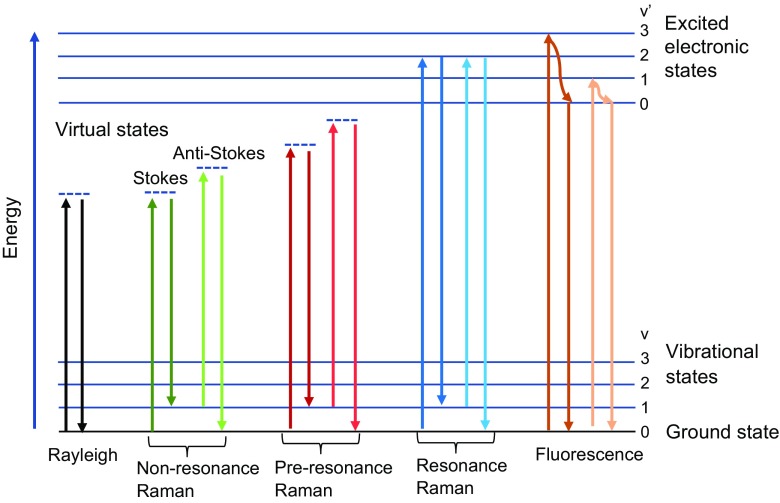
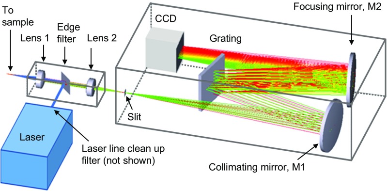
Novel approaches toward understanding the evolution of disease can lead to the discovery of biomarkers that will enable better management of disease progression and improve prognostic evaluation. Raman spectroscopy is a promising investigative and diagnostic tool that can assist in uncovering the molecular basis of disease and provide objective, quantifiable molecular information for diagnosis and treatment evaluation. This technique probes molecular vibrations/rotations associated with chemical bonds in a sample to obtain information on molecular structure, composition, and intermolecular interactions. Raman scattering occurs when light interacts with a molecular vibration/rotation and a change in polarizability takes place during molecular motion. This results in light being scattered at an optical frequency shifted (up or down) from the incident light. By monitoring the intensity profile of the inelastically scattered light as a function of frequency, the unique spectroscopic fingerprint of a tissue sample is obtained. Since each sample has a unique composition, the spectroscopic profile arising from Raman-active functional groups of nucleic acids, proteins, lipids, and carbohydrates allows for the evaluation, characterization, and discrimination of tissue type. This review provides an overview of the theory of Raman spectroscopy, instrumentation used for measurement, and variation of Raman spectroscopic techniques for clinical applications in cancer, including detection of brain, ovarian, breast, prostate, and pancreatic cancers and circulating tumor cells.
Keywords: Applications; Cancer; Clinical; Diagnosis; Raman spectroscopy; Spectroscopy.
Figures


References
-
- Atkins PW. Physical chemistry. 5. New York: W. H. Freeman; 1994.
-
- Hecht E. Optics. 3. New York: Addison-Wesley; 1998.
-
- Raman CV. A new radiation. Indian Journal of Physics. 1928;2:387–398.
-
- Chase B. A new generation of Raman instrumentation. Applied Spectroscopy. 1994;48(7):14A–19A.
-
- Bernath PF. Light scattering and the Raman effect. In: Bernath PF, editor. Spectra of atoms and molecules. 2. New York: Oxford University Press Inc; 2005. pp. 293–317.
Publication types
MeSH terms
LinkOut - more resources
Full Text Sources
Other Literature Sources
